Electron micrographs used with permission from the University of Texas Southwestern Medical School.
| Click on the image you would like to view: | |||||
|
Pancreas 1
|
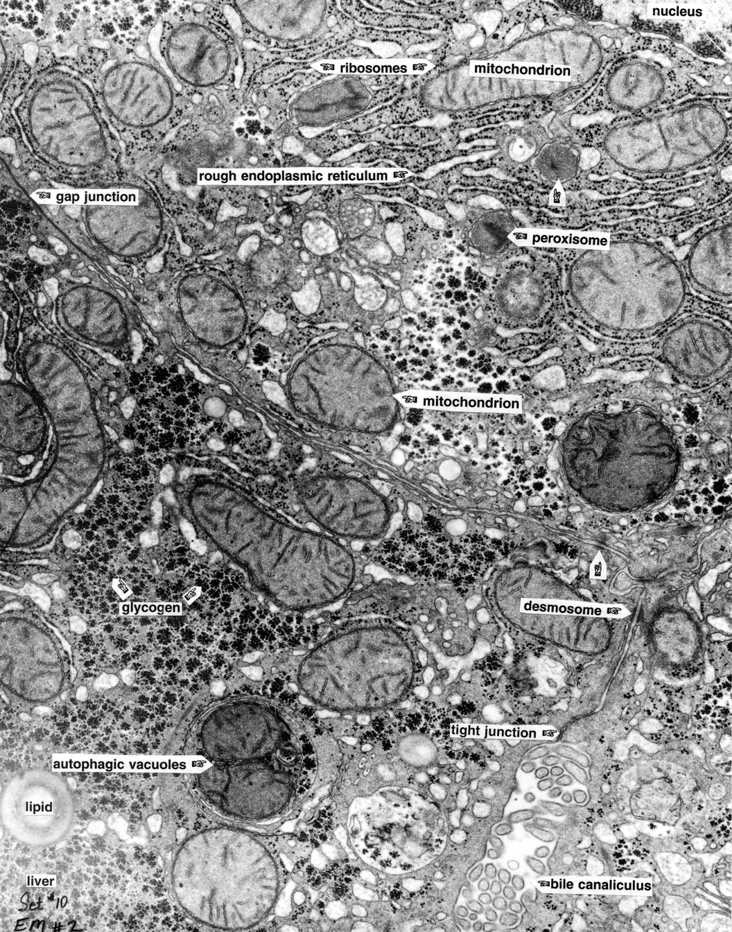
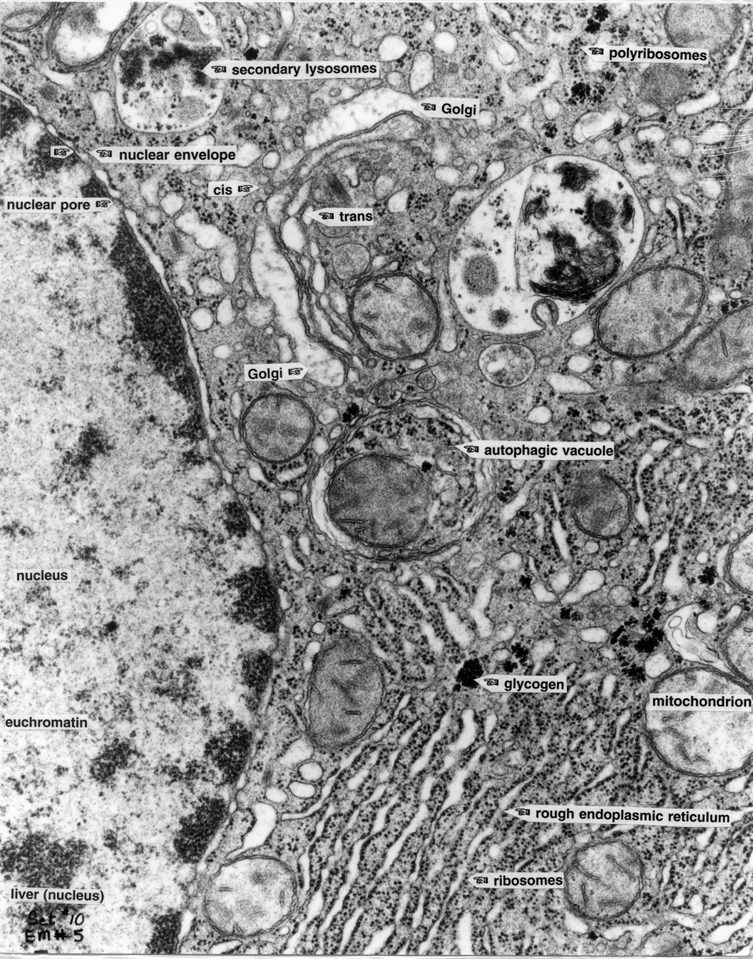
Liver 2 |
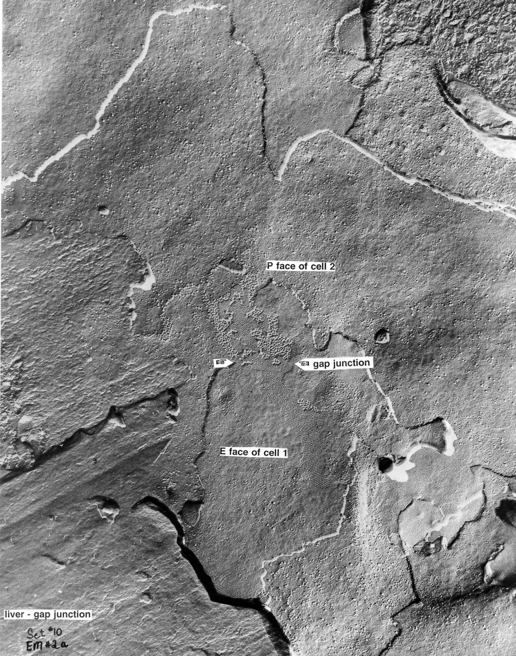
Liver 2a |
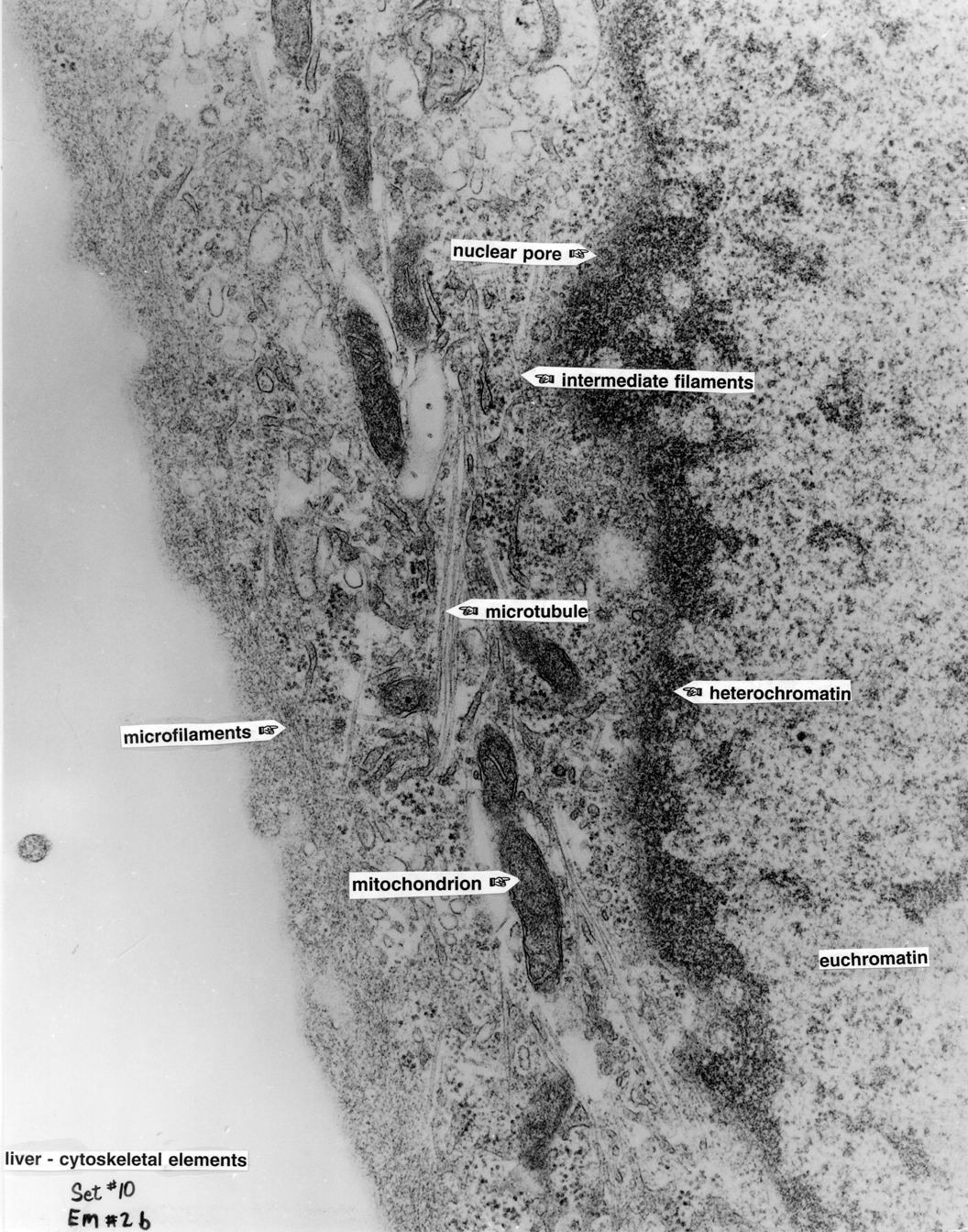
Liver 2b |
Gap Junction 3 |
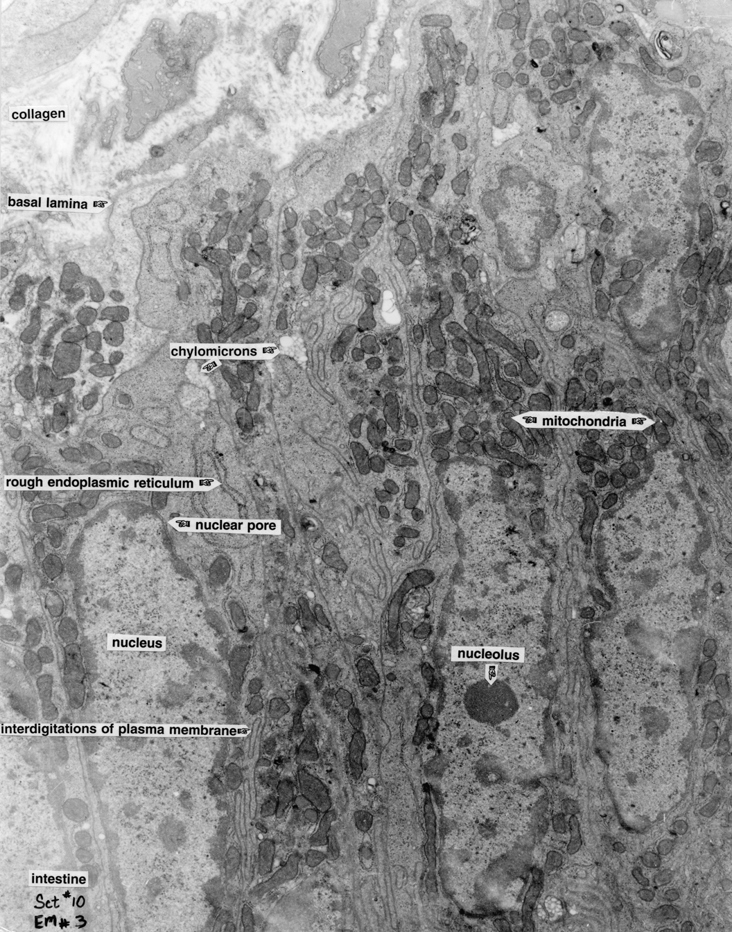
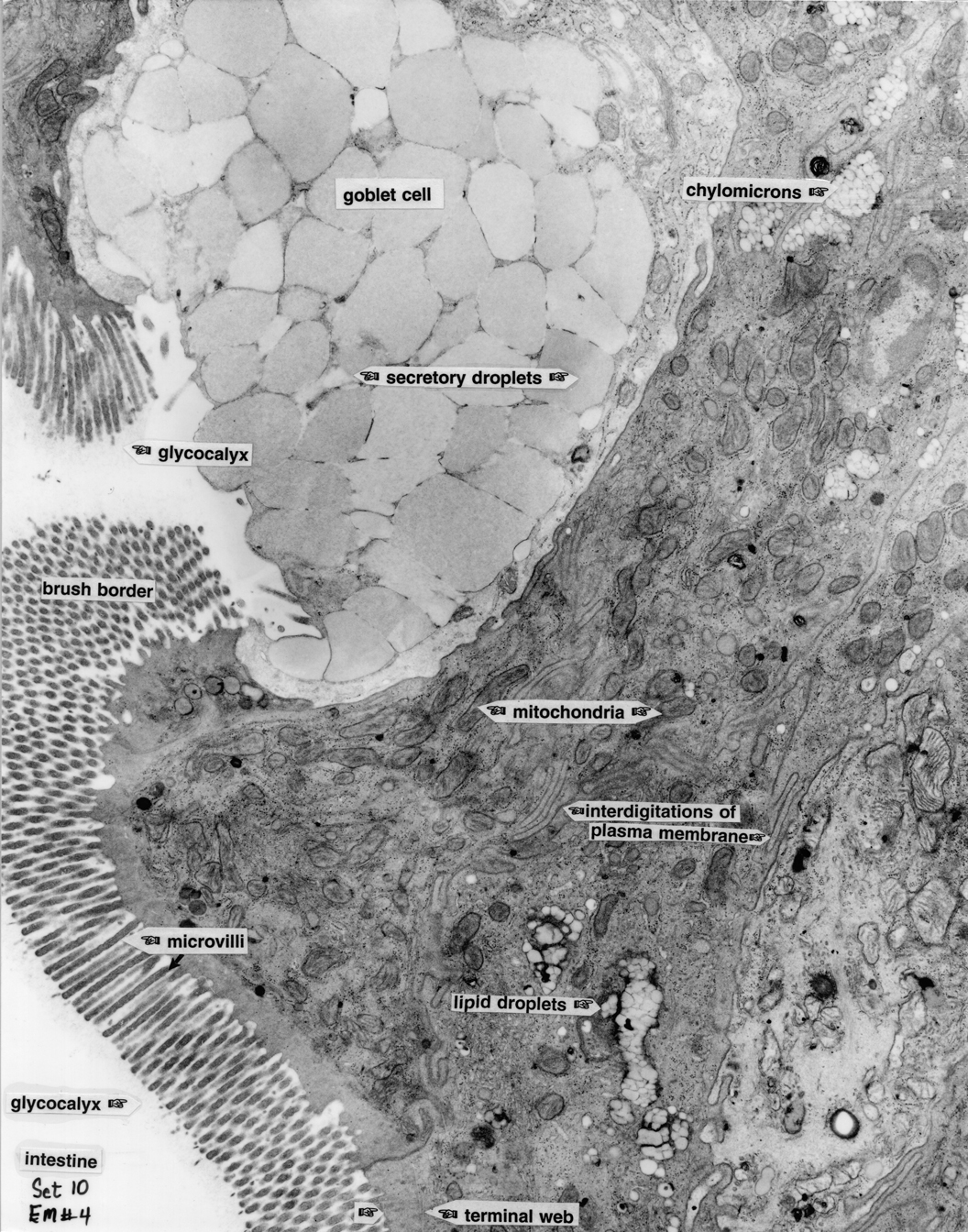
Intestine 4 |
|
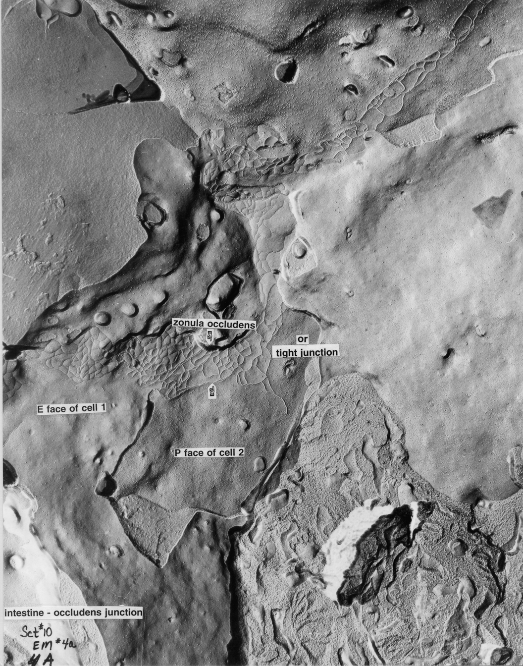
Intestine 4a |
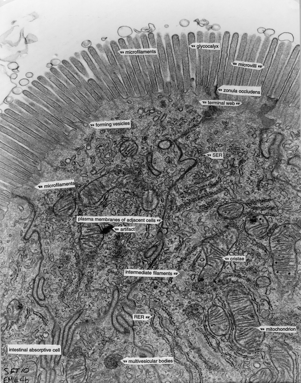
Intestine 4b |
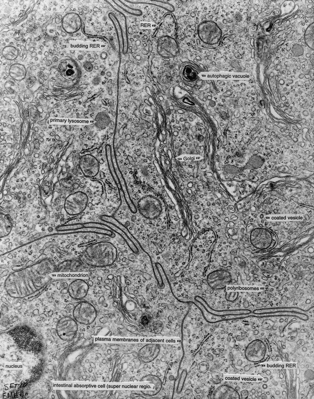
Intestine 4c |
Liver 5 |
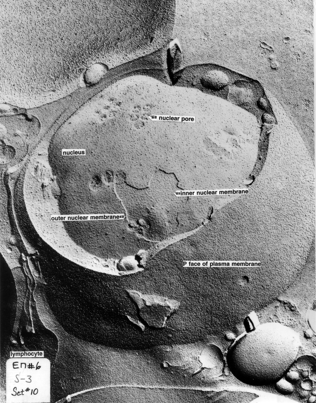
Lymphocyte 6 |
Centriole-Microtubules |
|
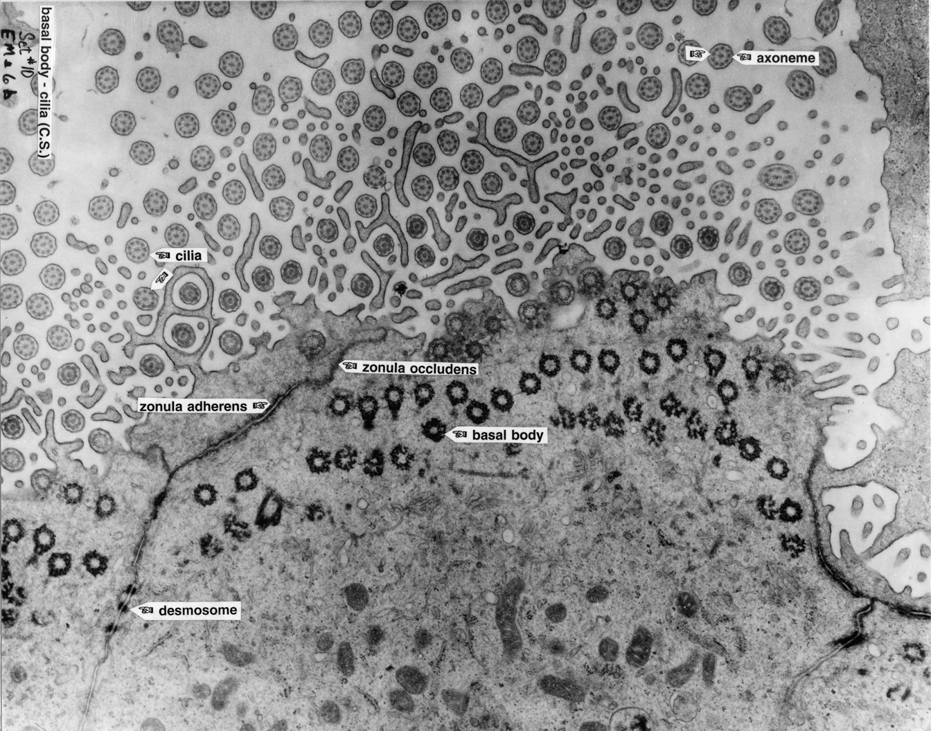
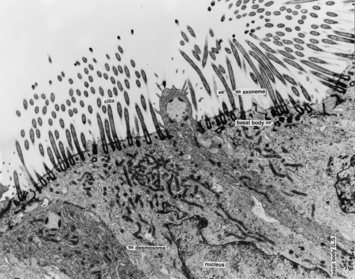
Ciliated Cells 6b |
Ciliated Cells 6c |
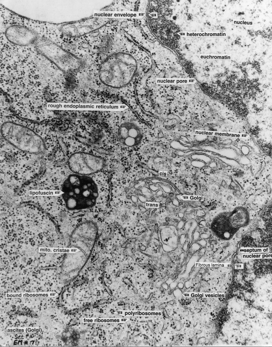
Cell Cytoplasm 7 |
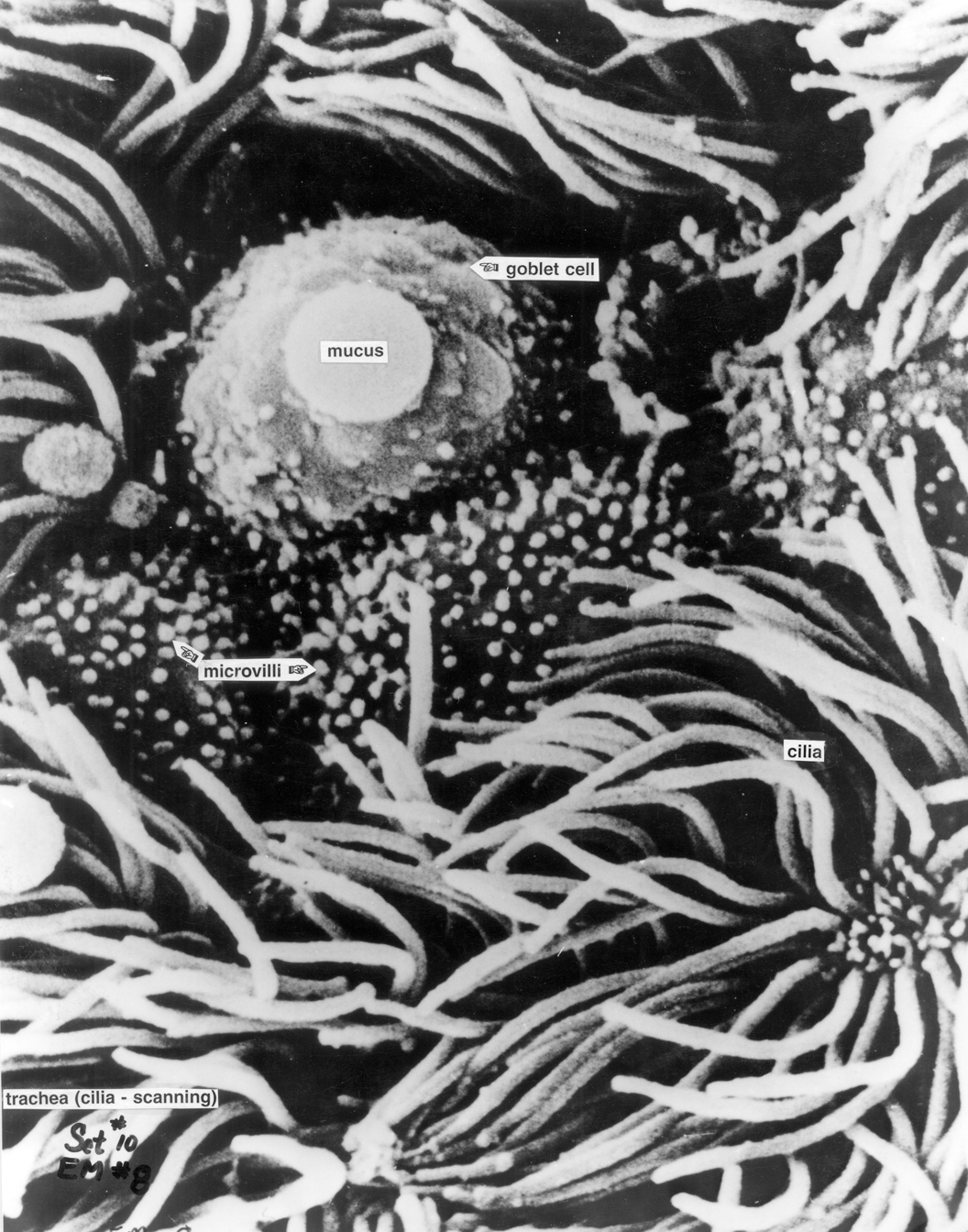
Trachea |
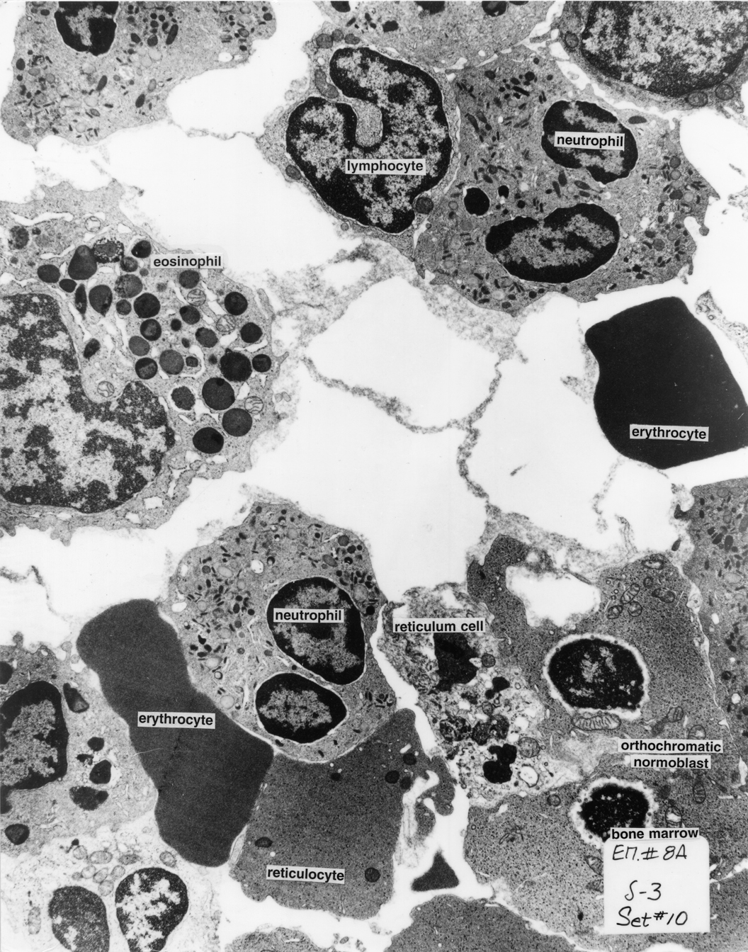
Bone Marrow |
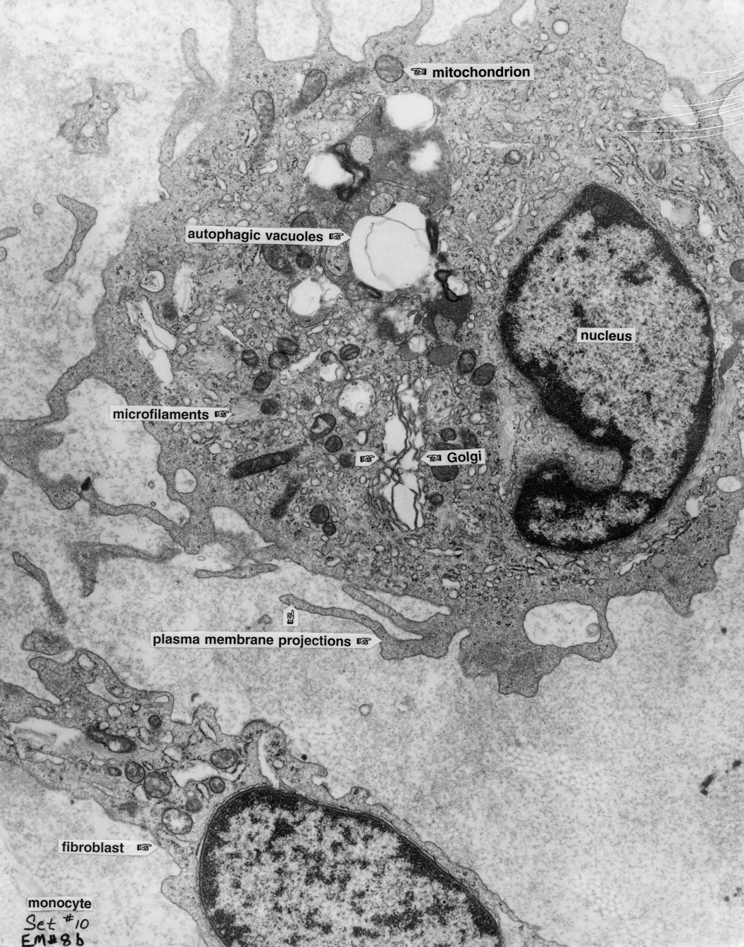
Monocyte 8b |
|
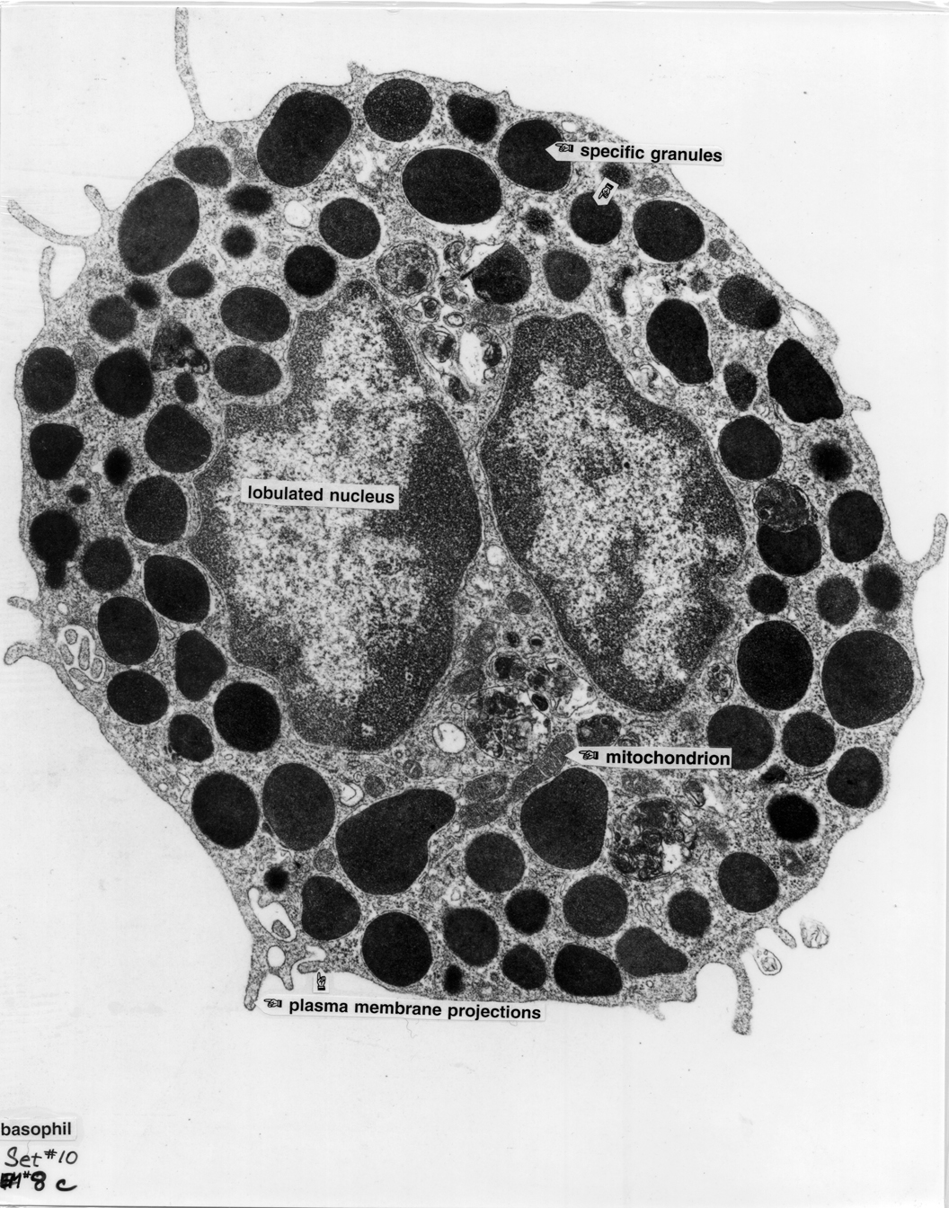
Basophil 8c |
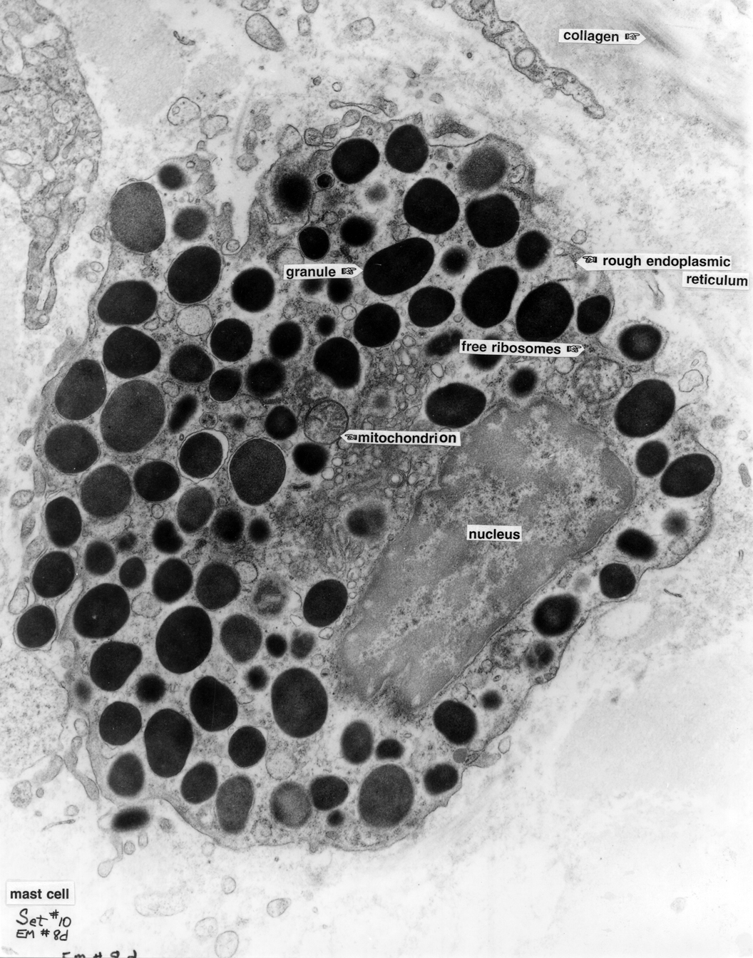
Mast Cell 8d |
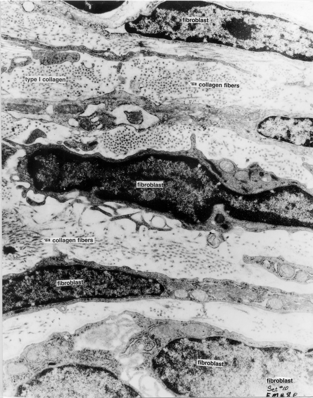
Fibroblast 8e |
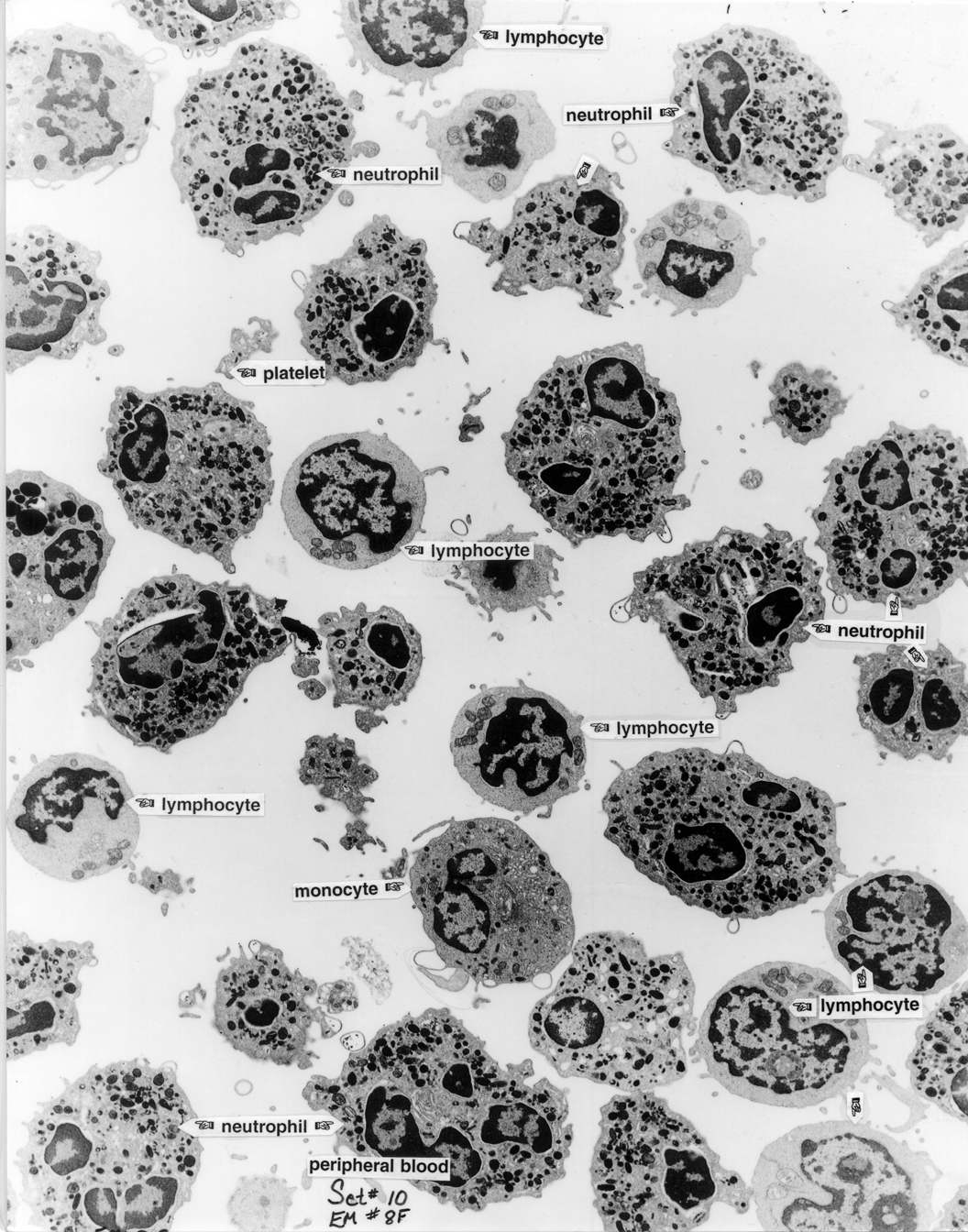
Peripheral Blood 8f |
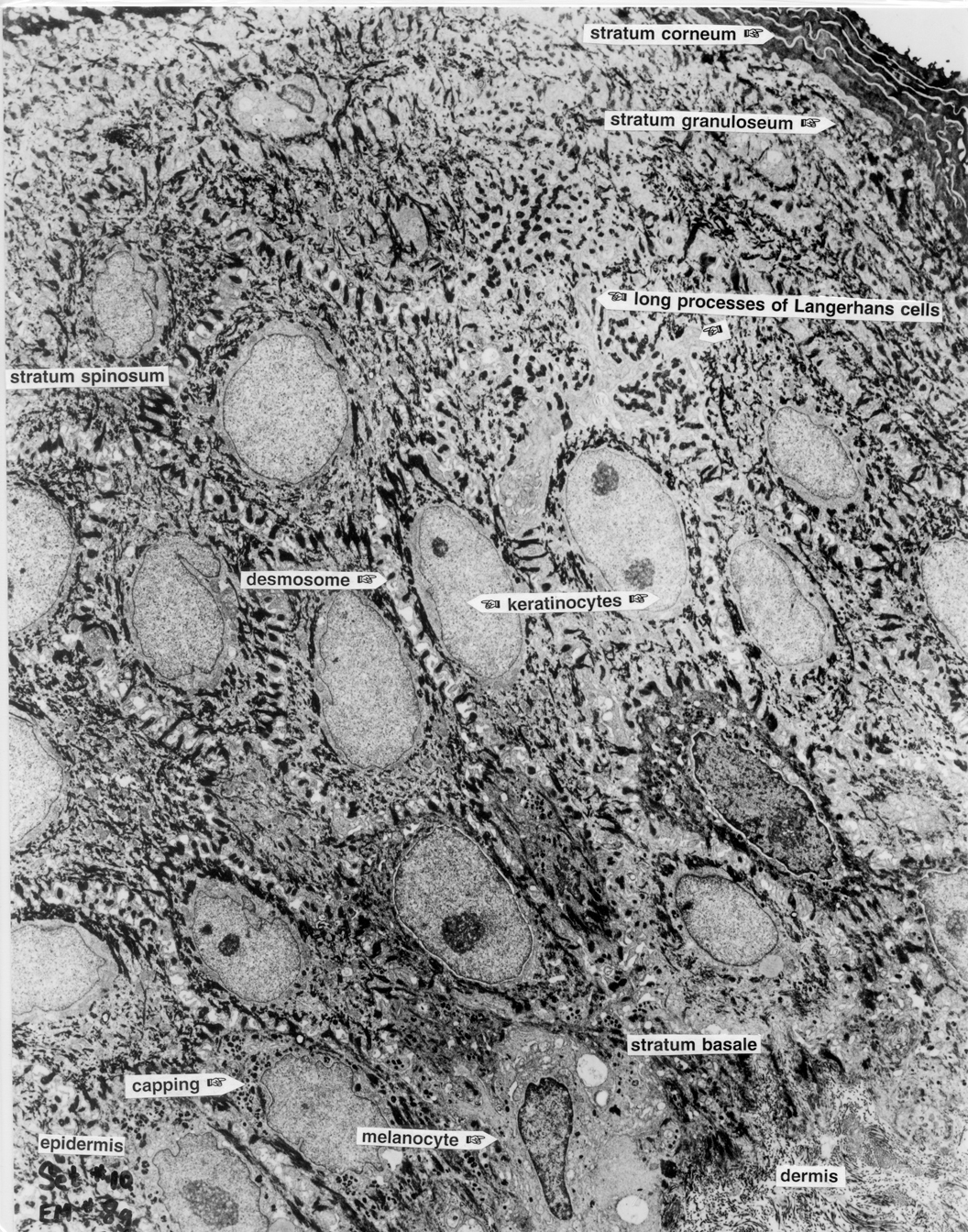
Epidermis 8g |
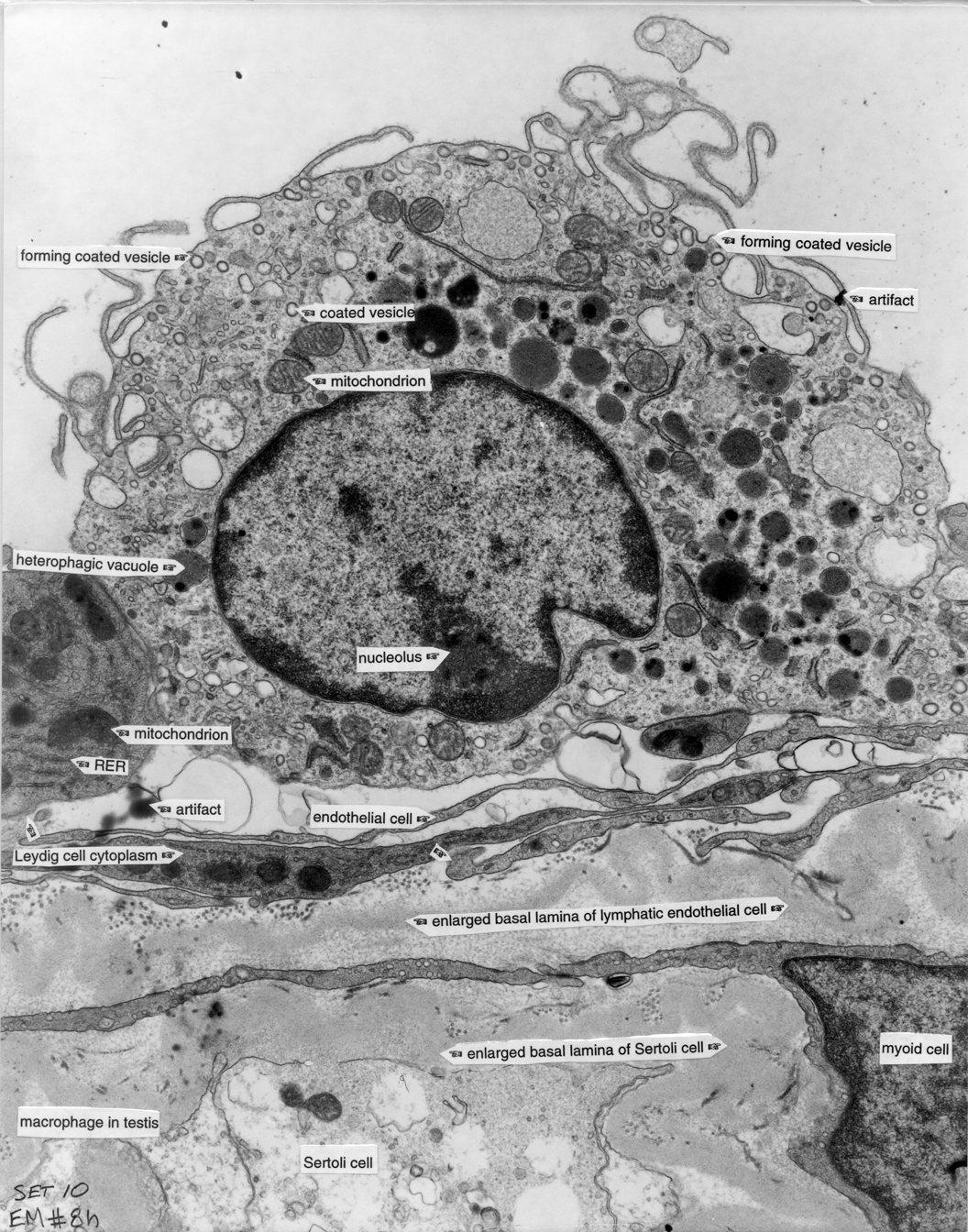
Macrophage 8h |
|
Smooth Muscle 9 |
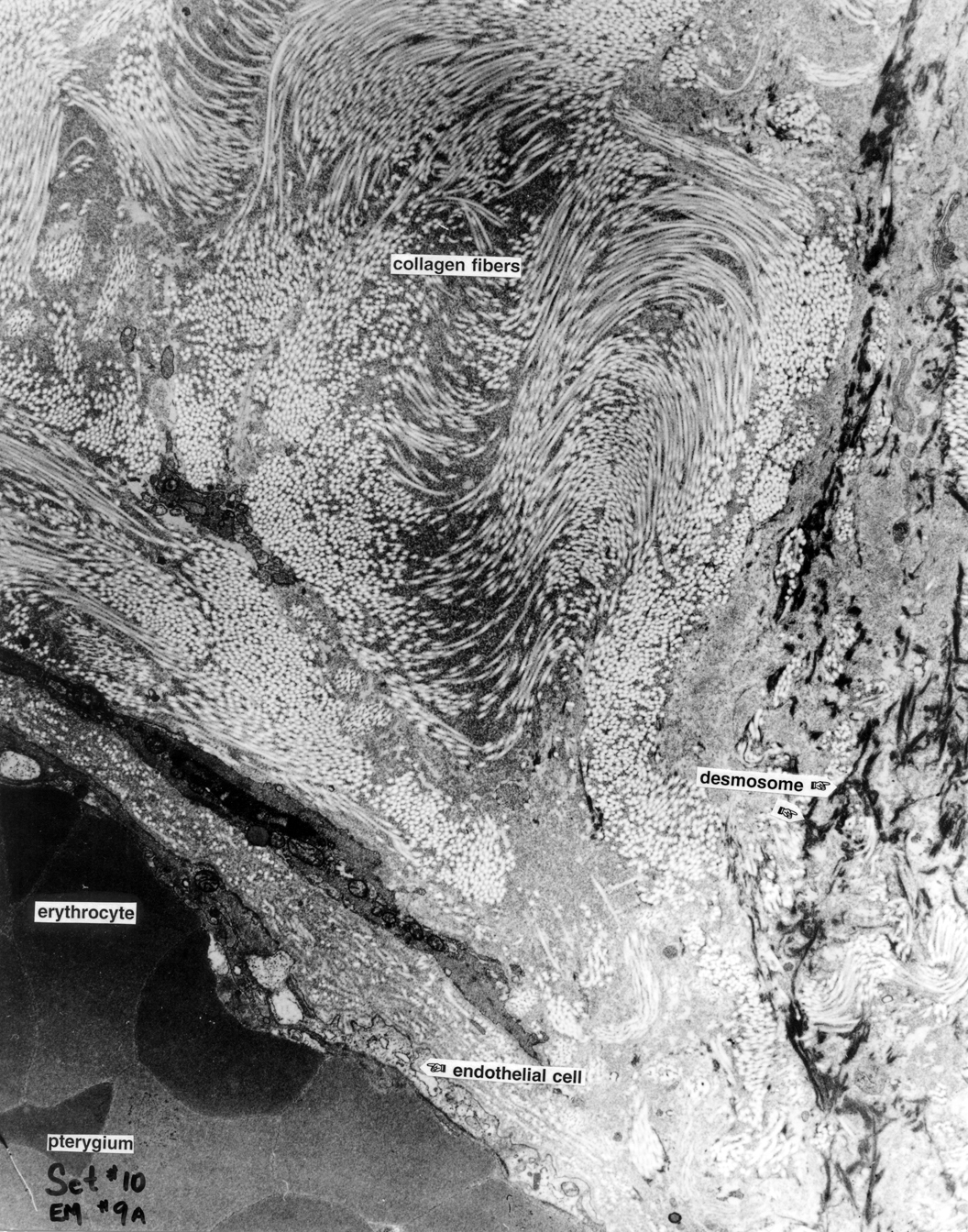
Skin 9a |
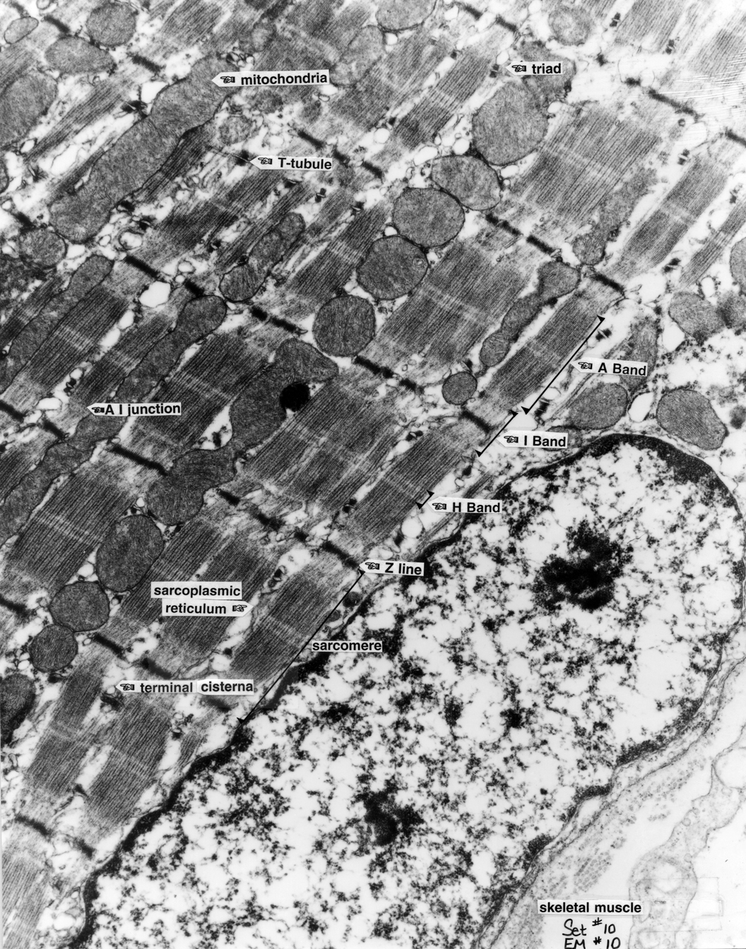
Skeletal Muscle 10 |
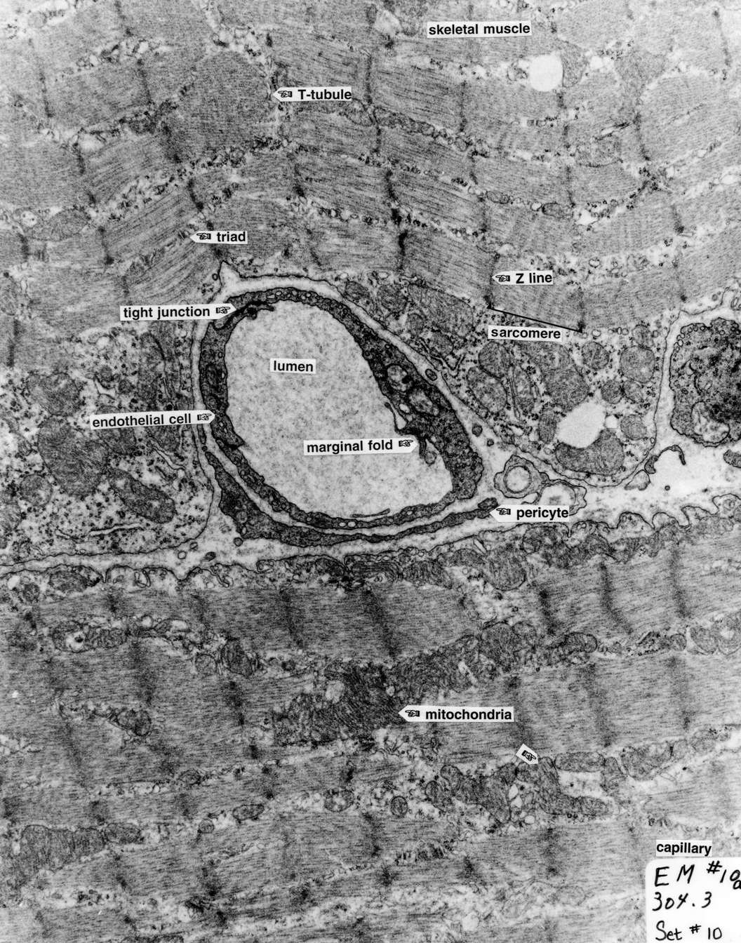
Capillary 10a |
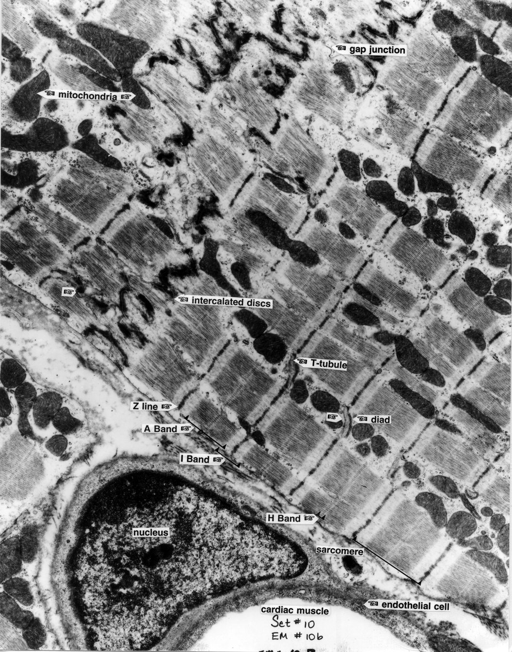
Cardiac Muscle |
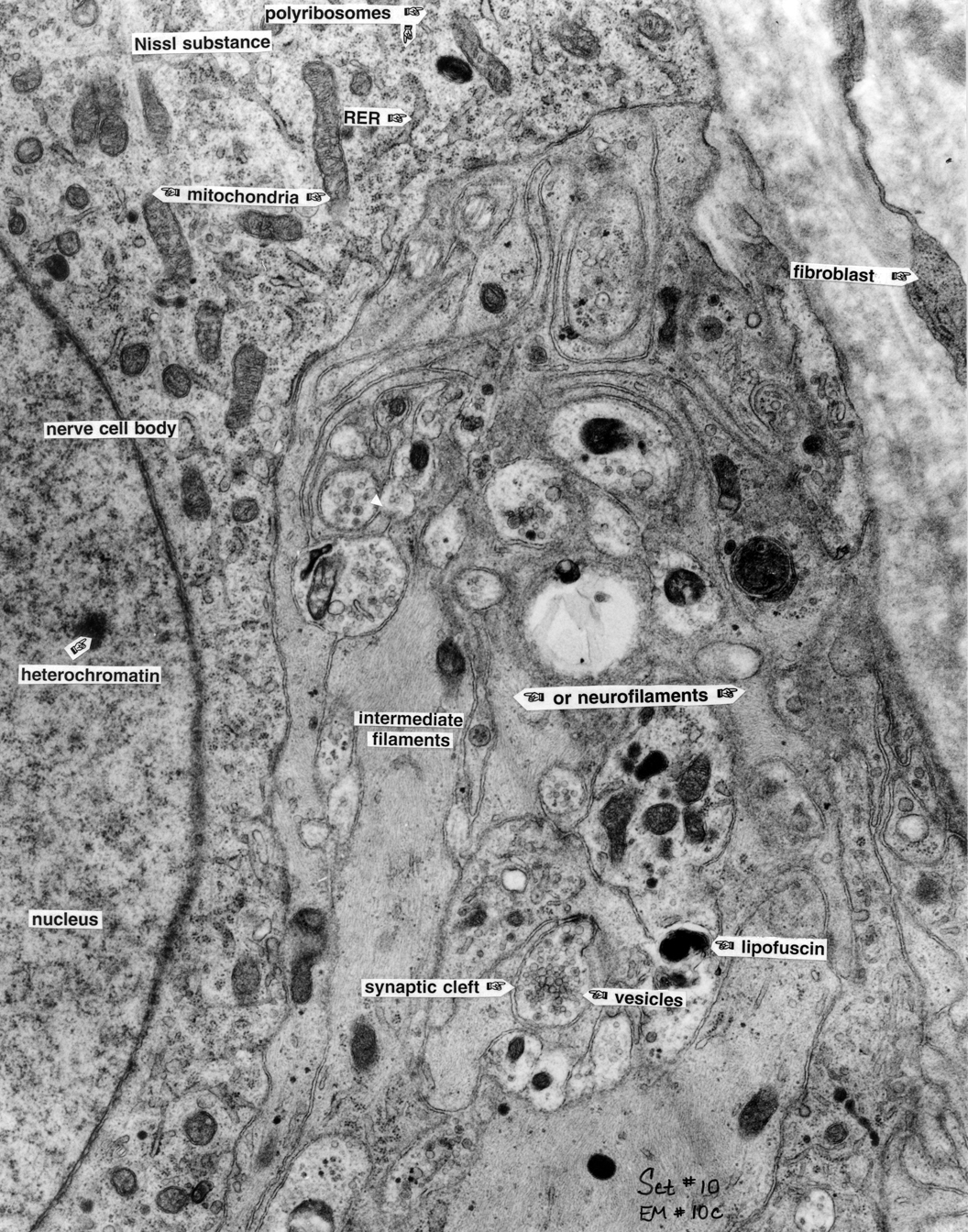
Nerve Cell Body 10c |
|
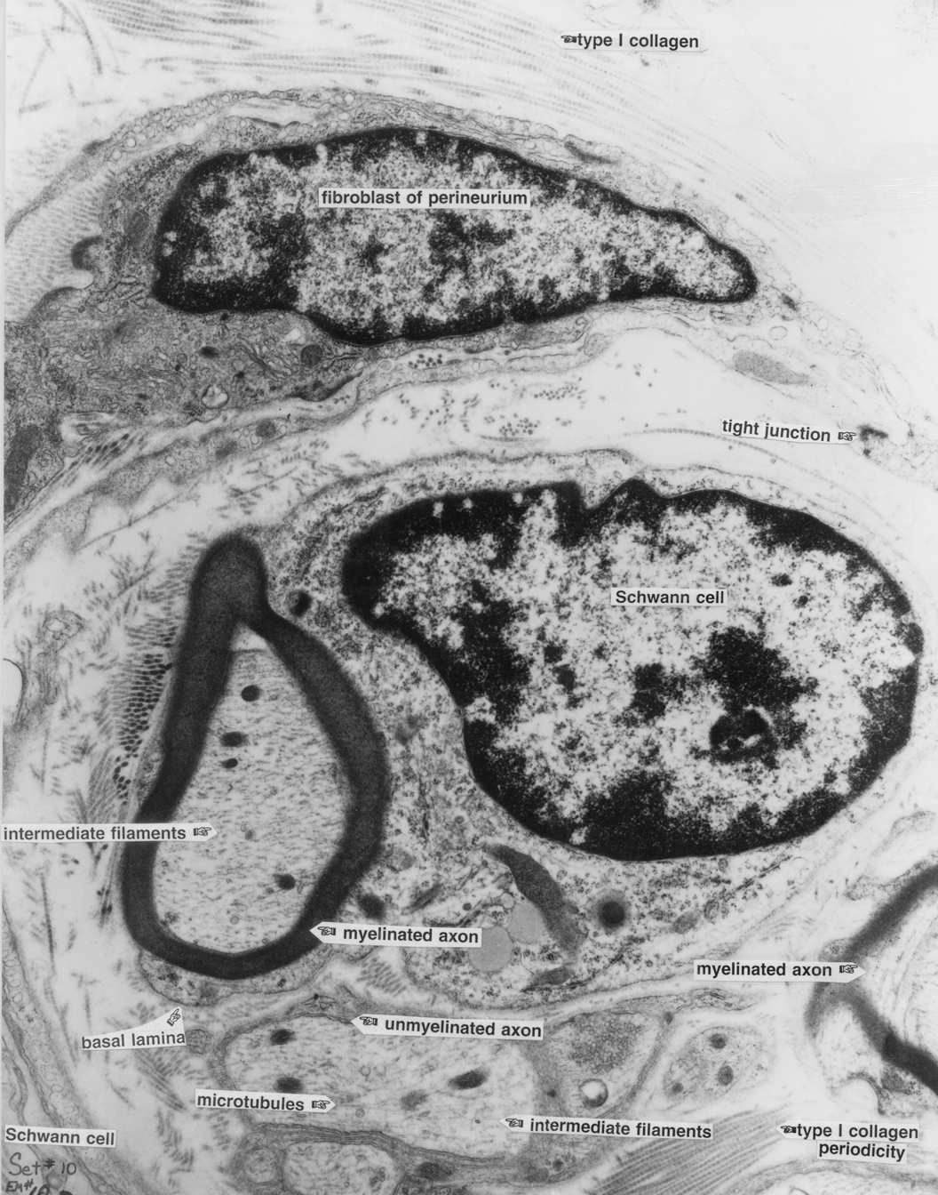
Schwann Cell of Nerve 10d |
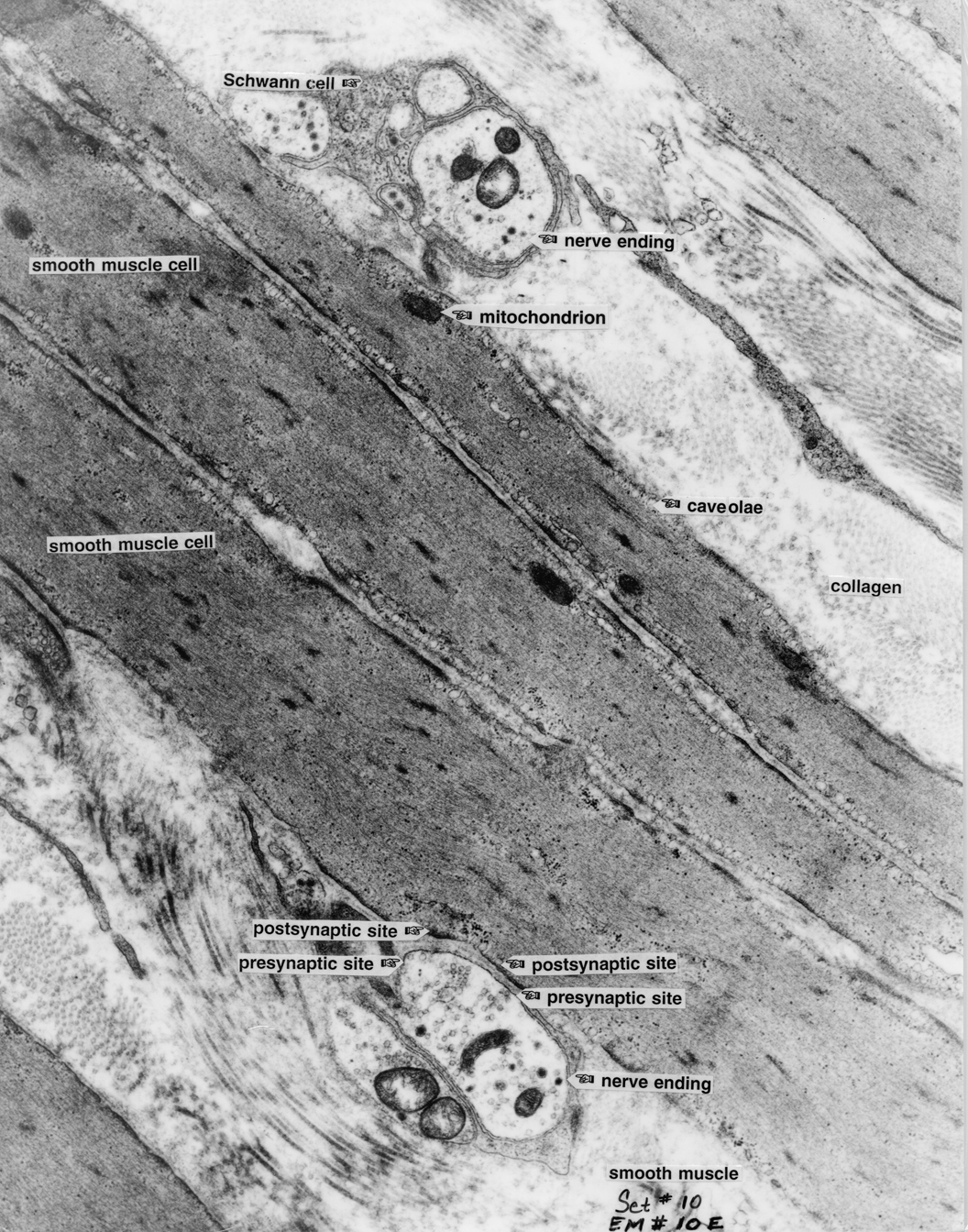
Smooth Muscle 10e |
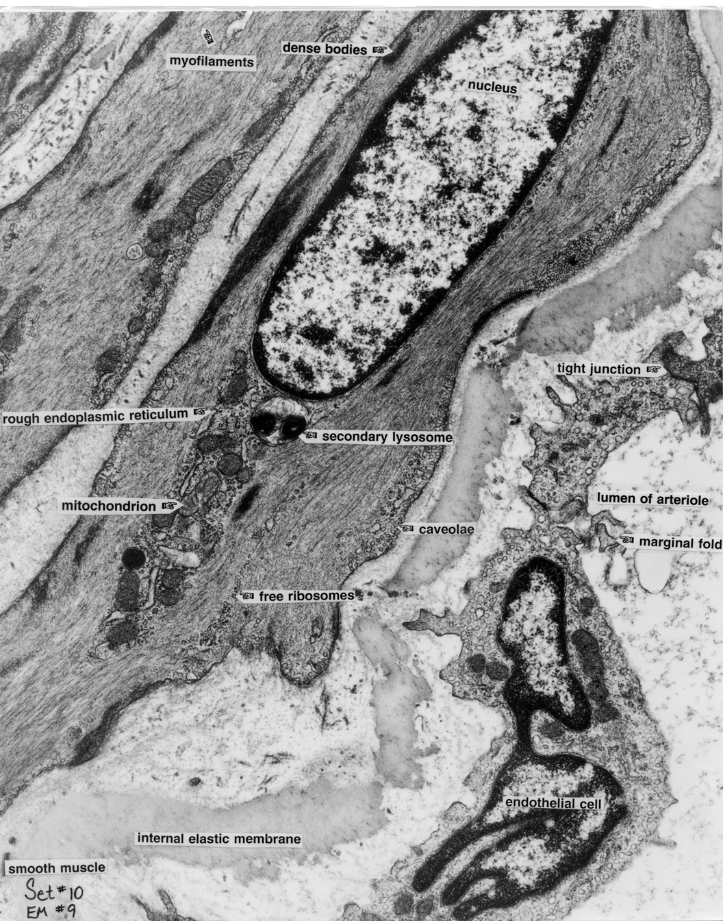
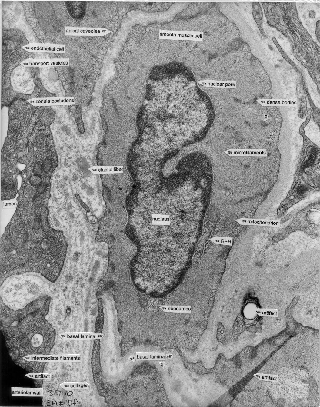
Arteriole 10f |
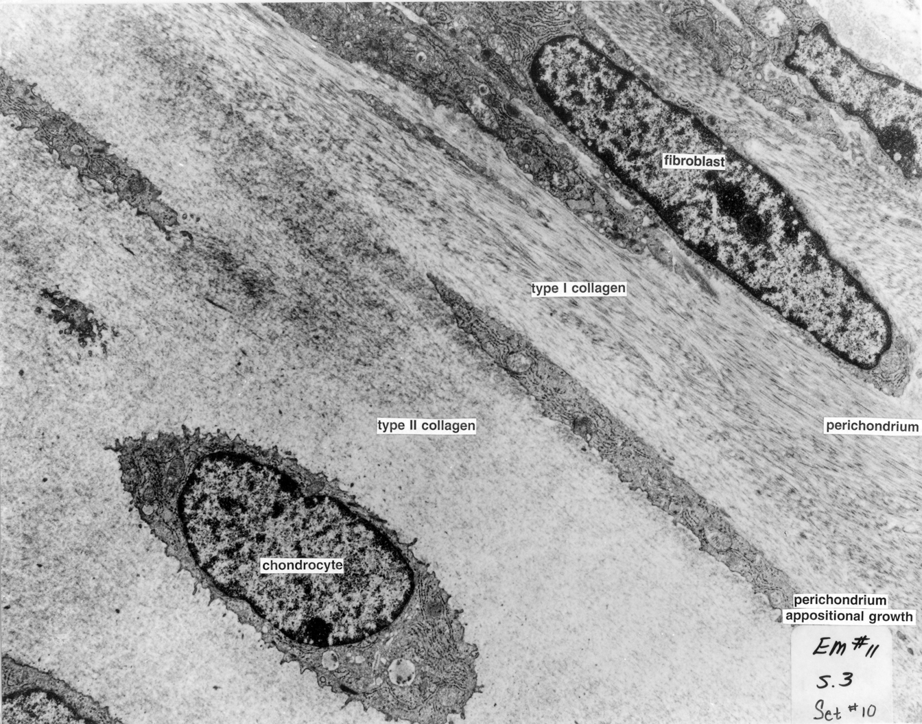
Perichondrium of Cartilage 11 |
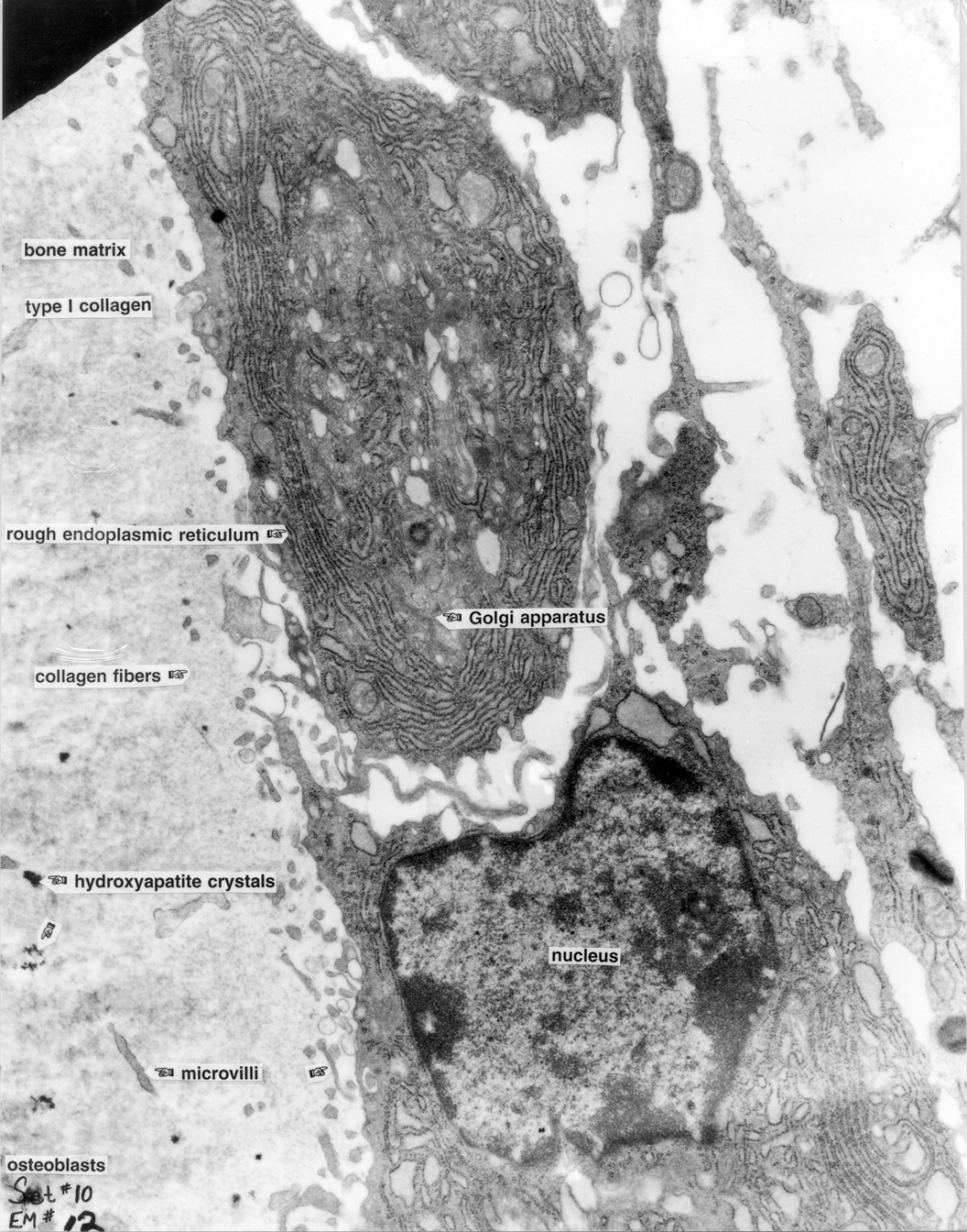
Osteoblasts 12 |
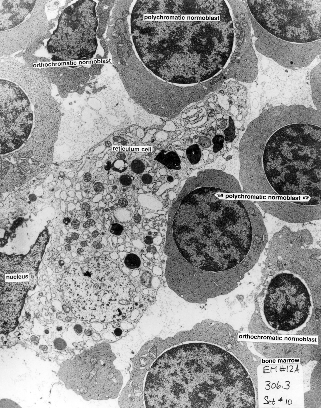
Bone Marrow 12a |
|
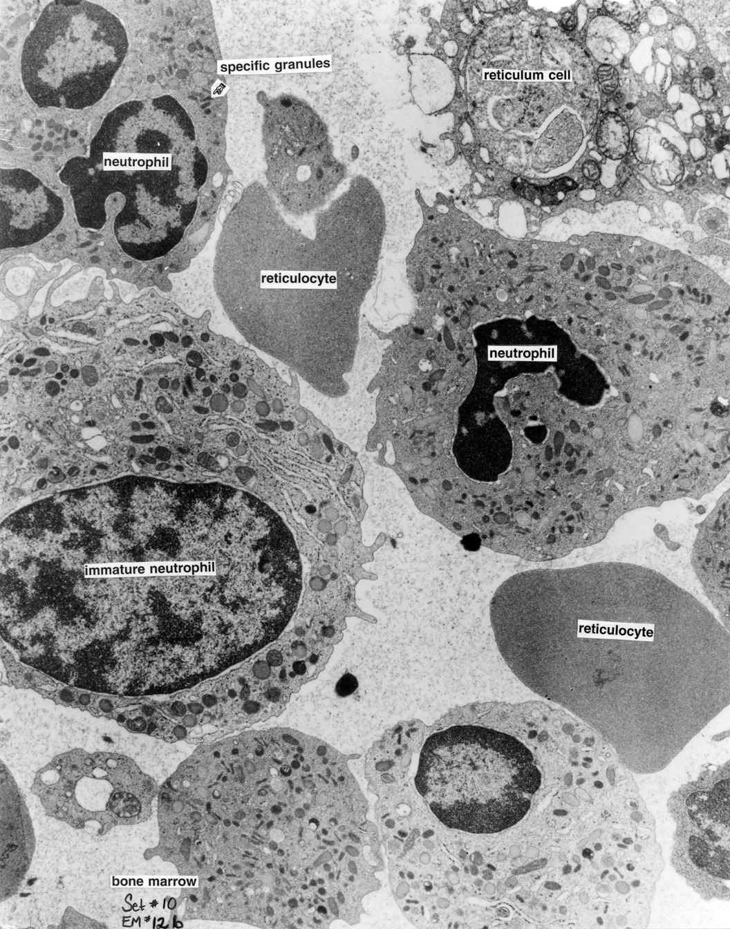
Bone Marrow 12b |
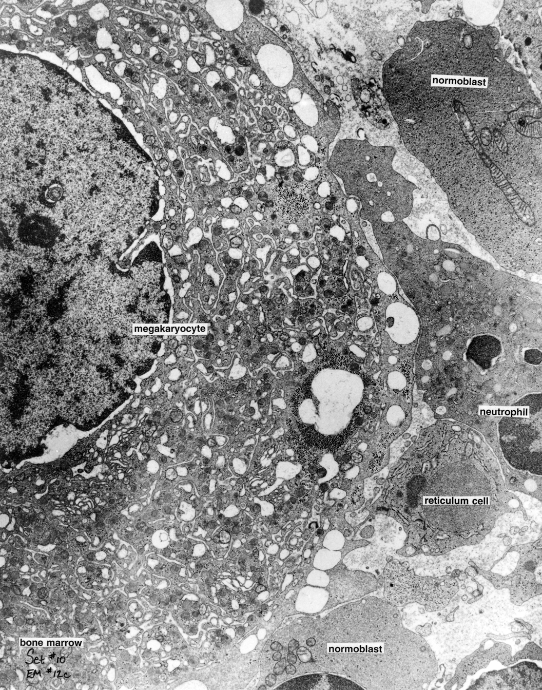
Bone Marrow 12c |
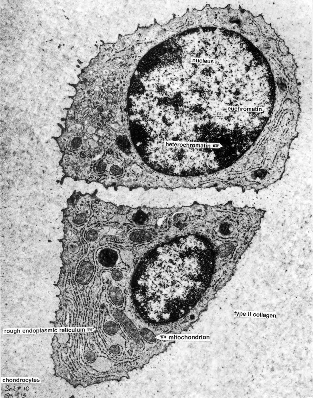
Cartilage 13 |
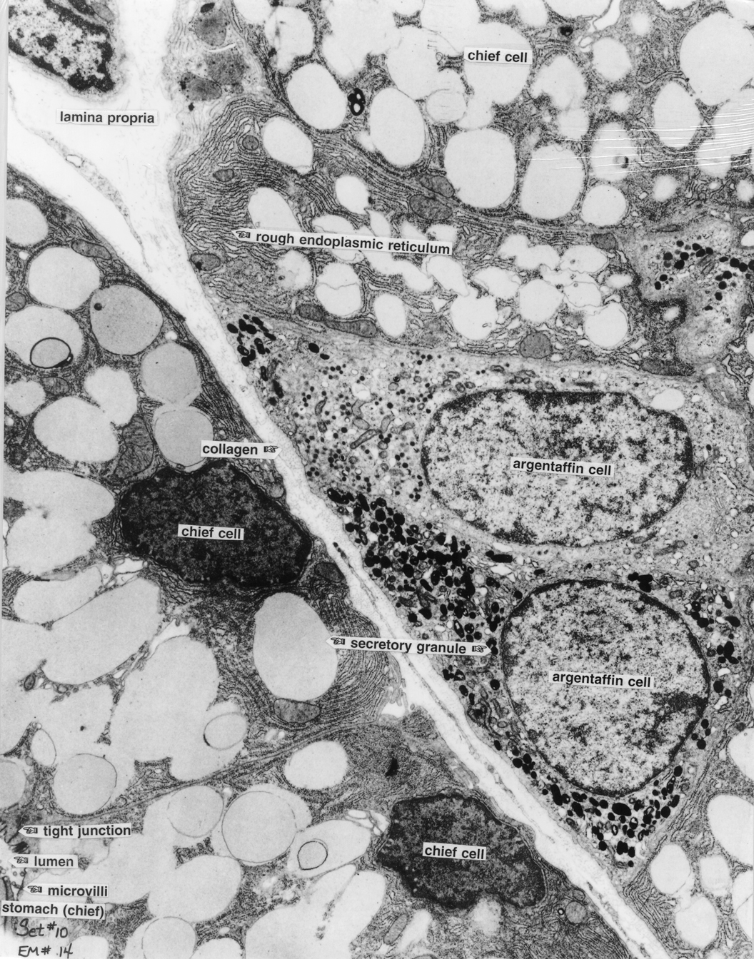
Stomach 14 |
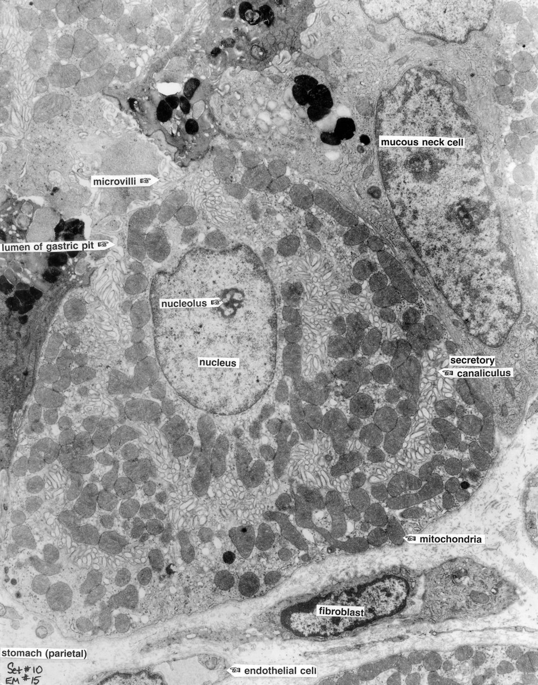
Stomach 15 |
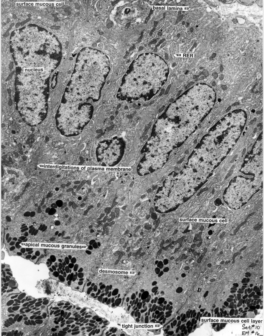
Stomach 16 |
|
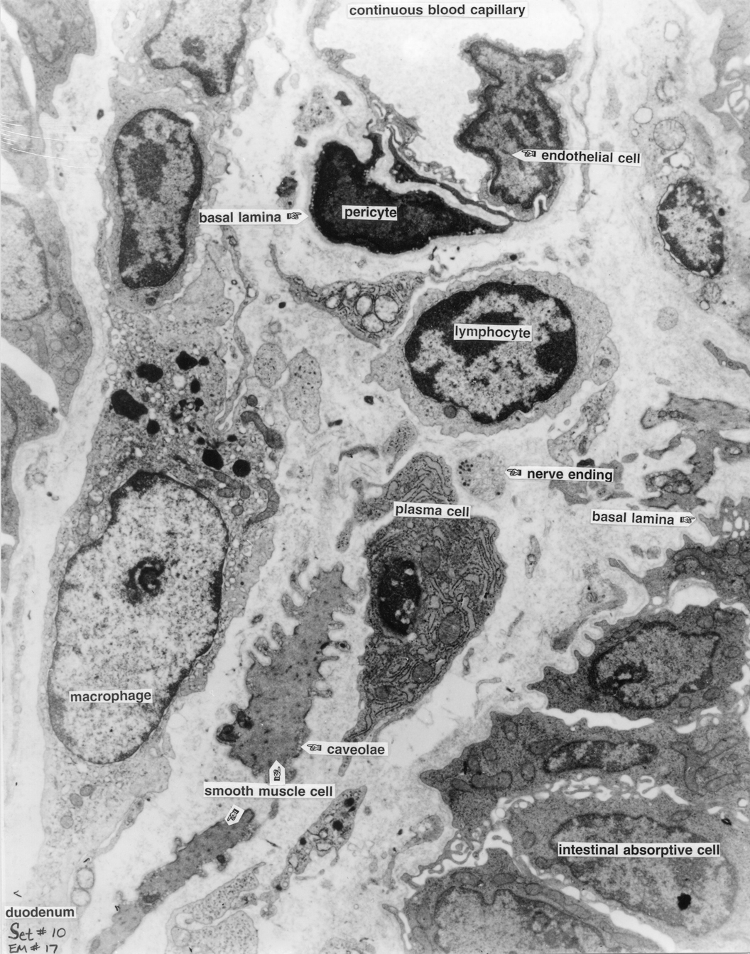
Intestine 17 |
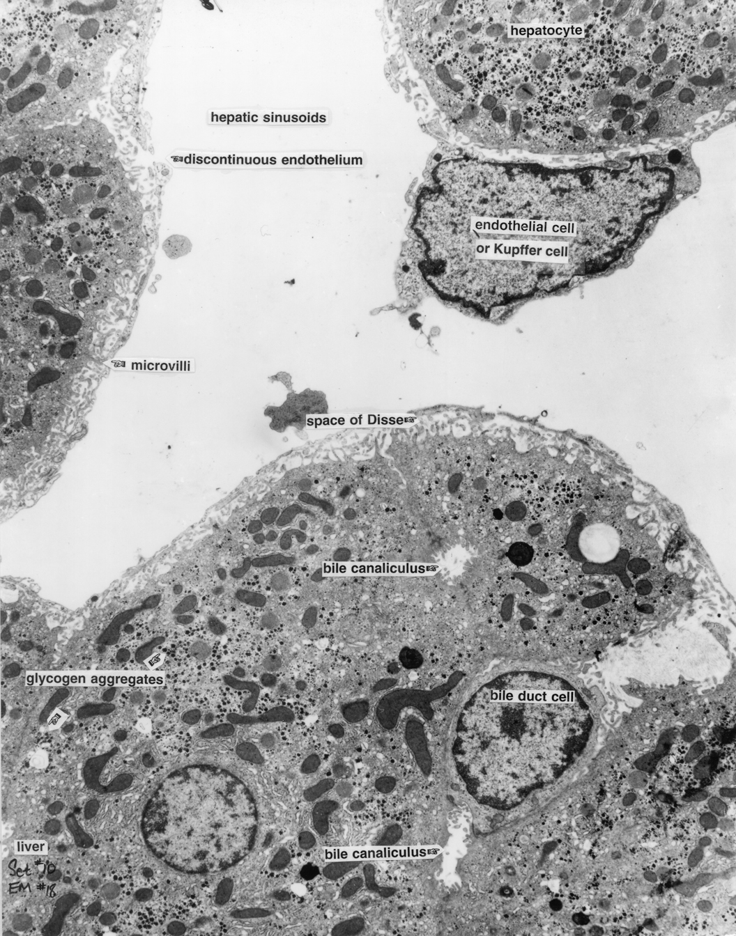
Liver 18 |
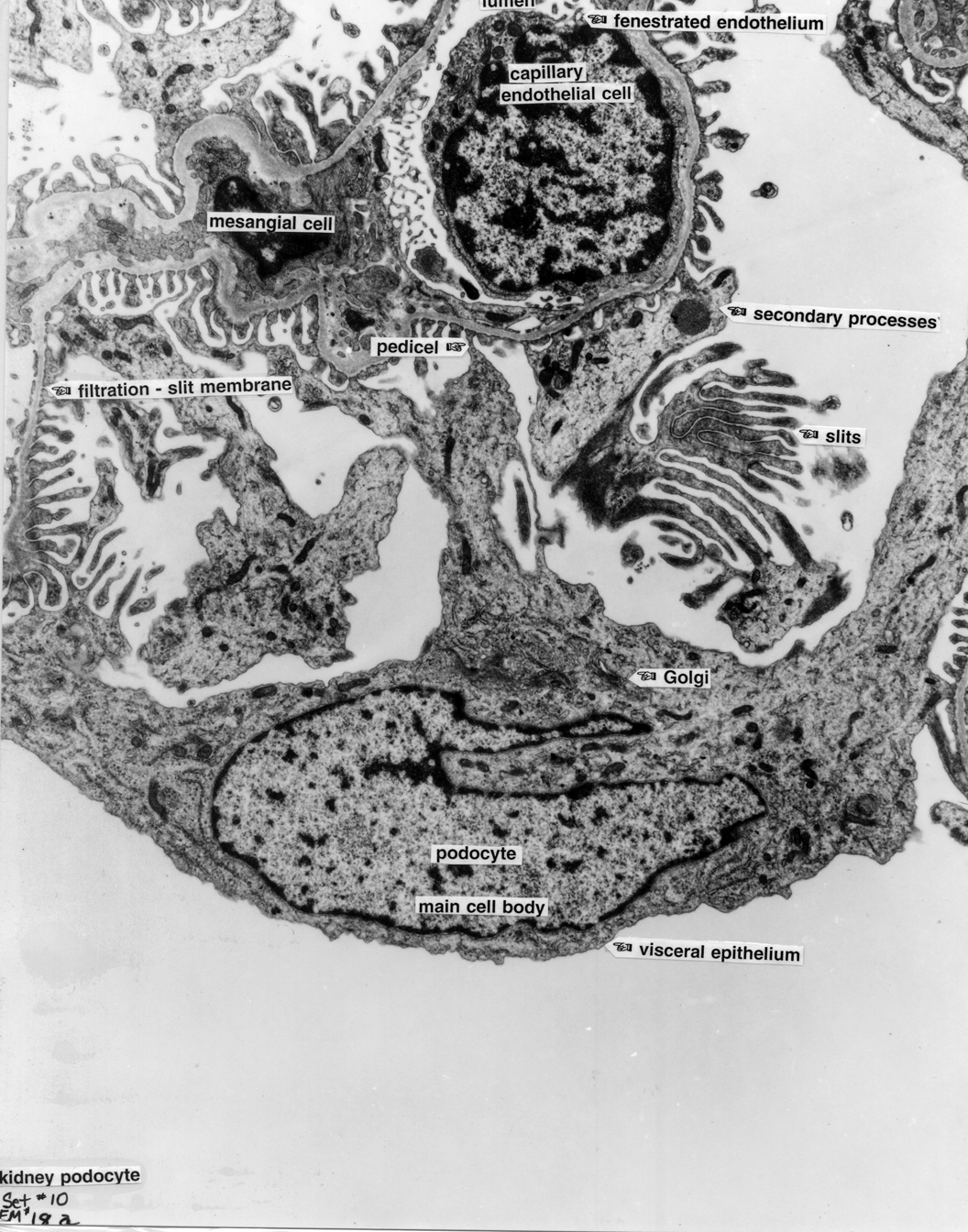
Kidney 18a |
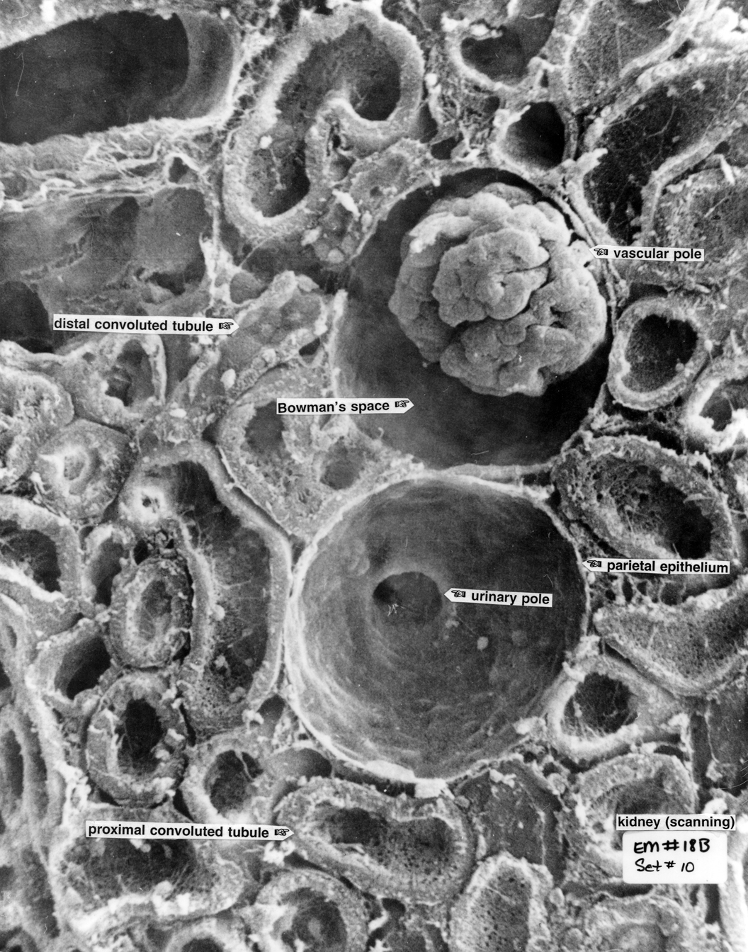
Kidney 18b |
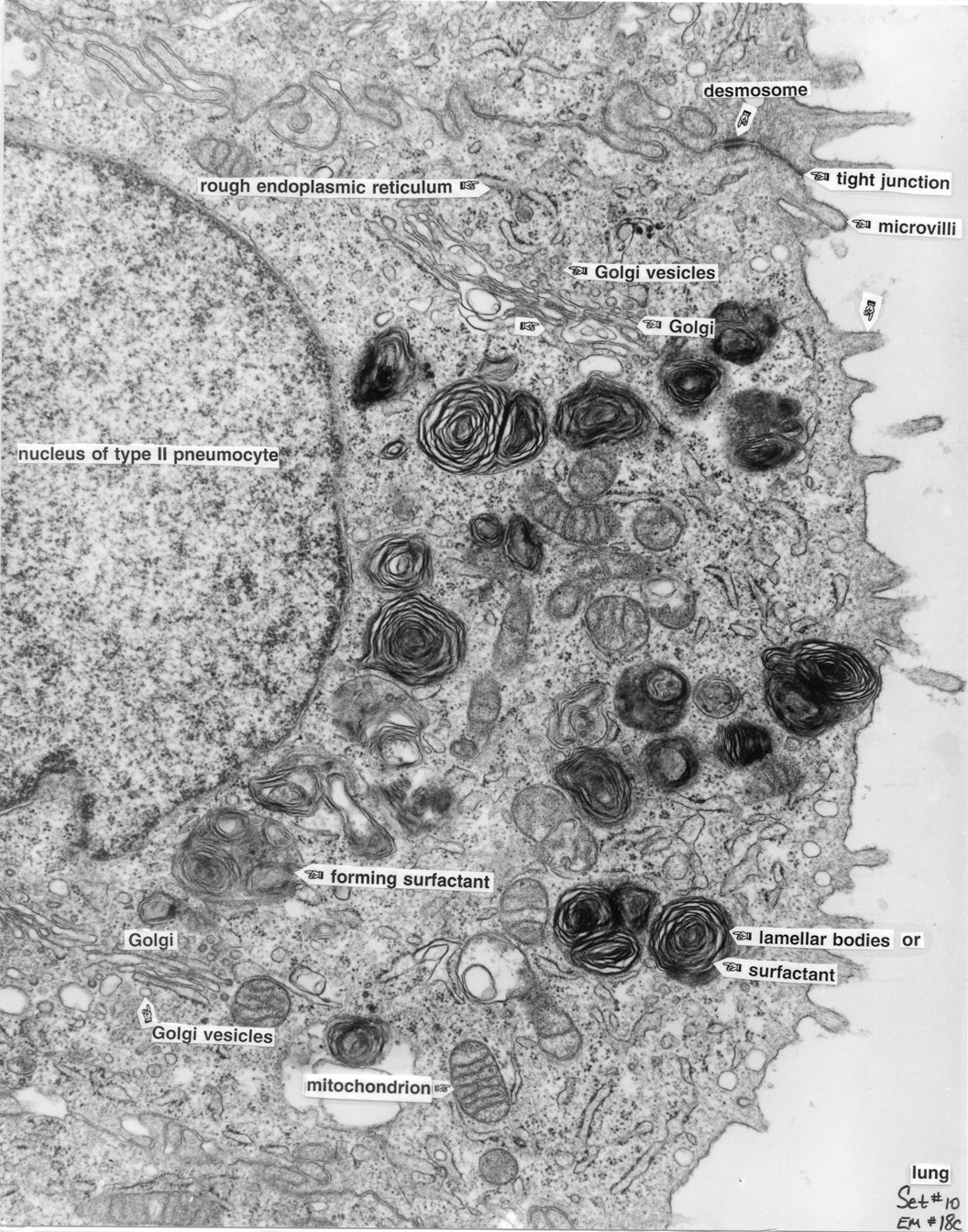
Lung 18c |
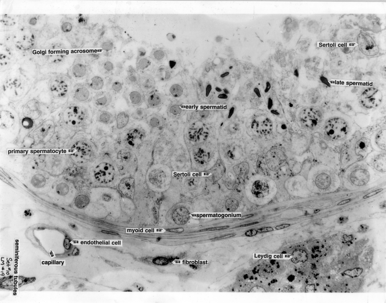
Testis 19 |
|
Testis 19a |
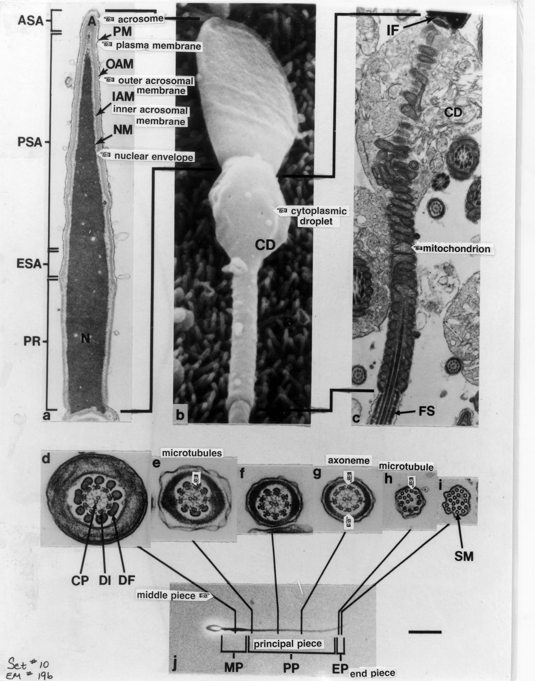
Speratozoa 19b |
Sertoli Cell and Germ Cells 19c |
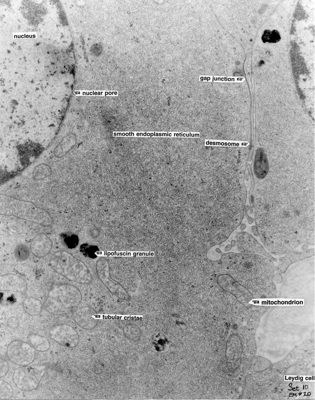
Leydig Cell 20 |
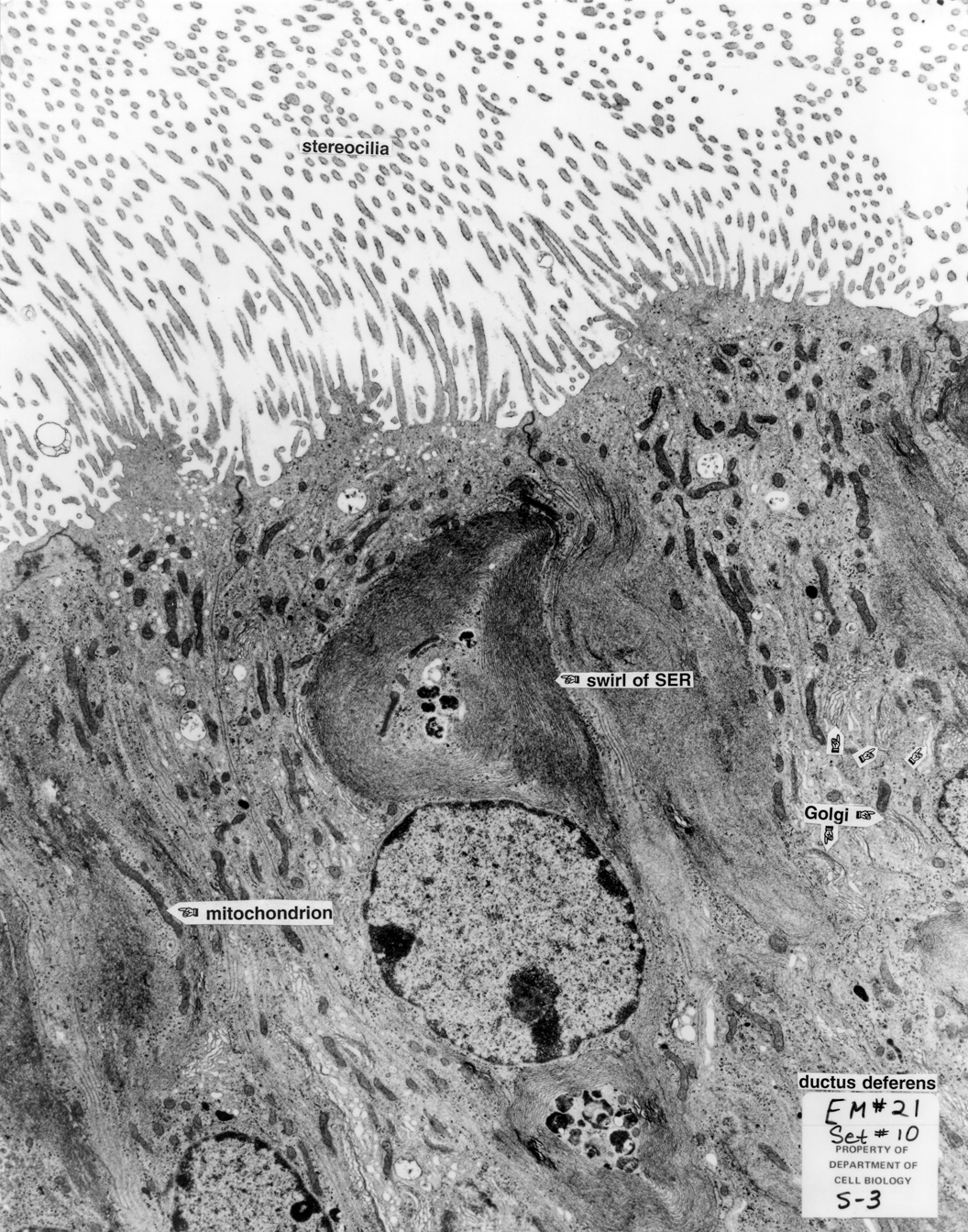
Ductus Deferens 21 |
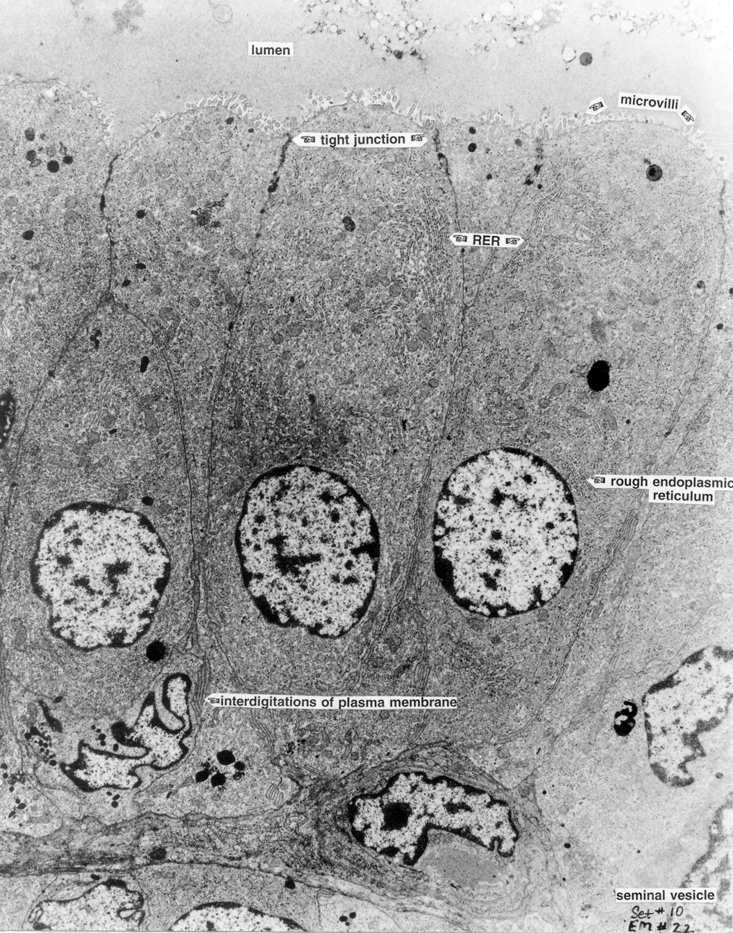
Seminal Vesicle 22 |
|
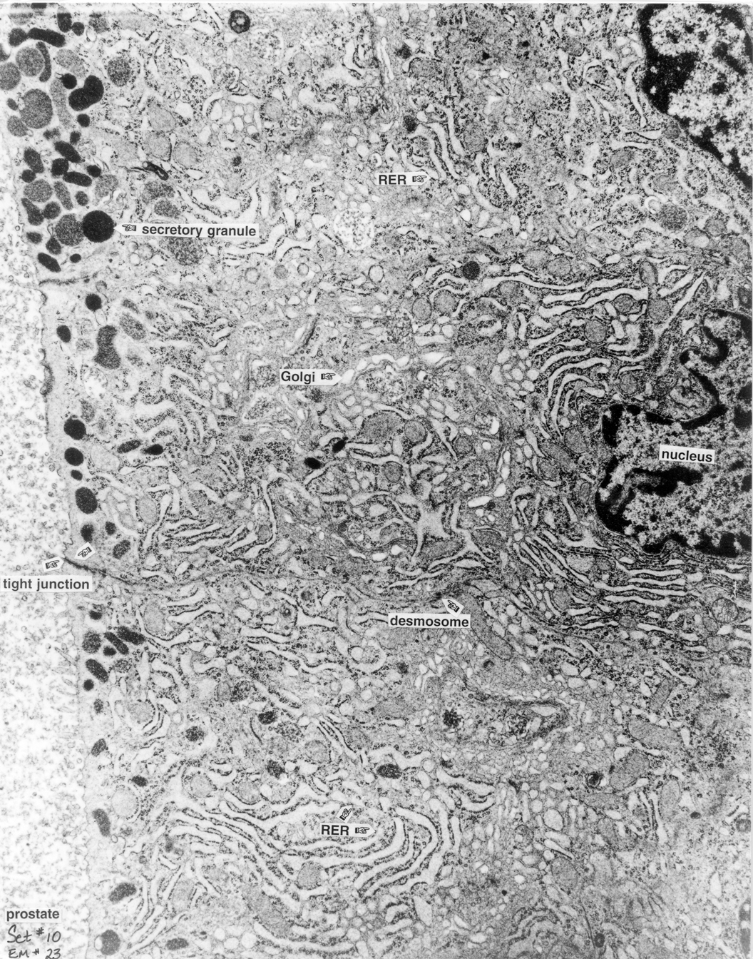
Prostate 23 |
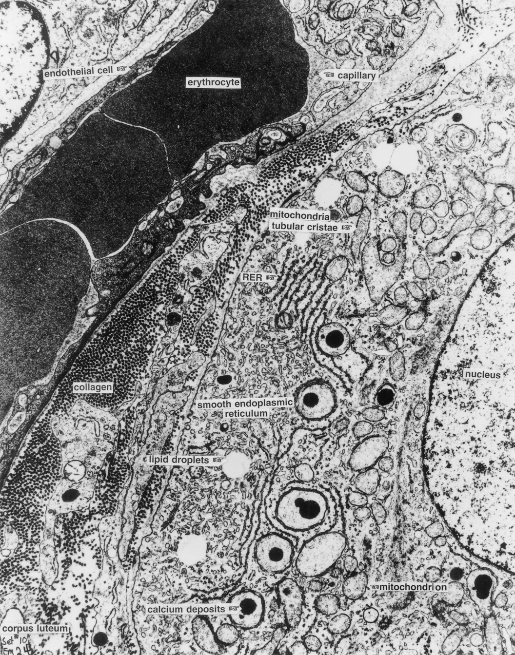
Corpus Luteum 24 |
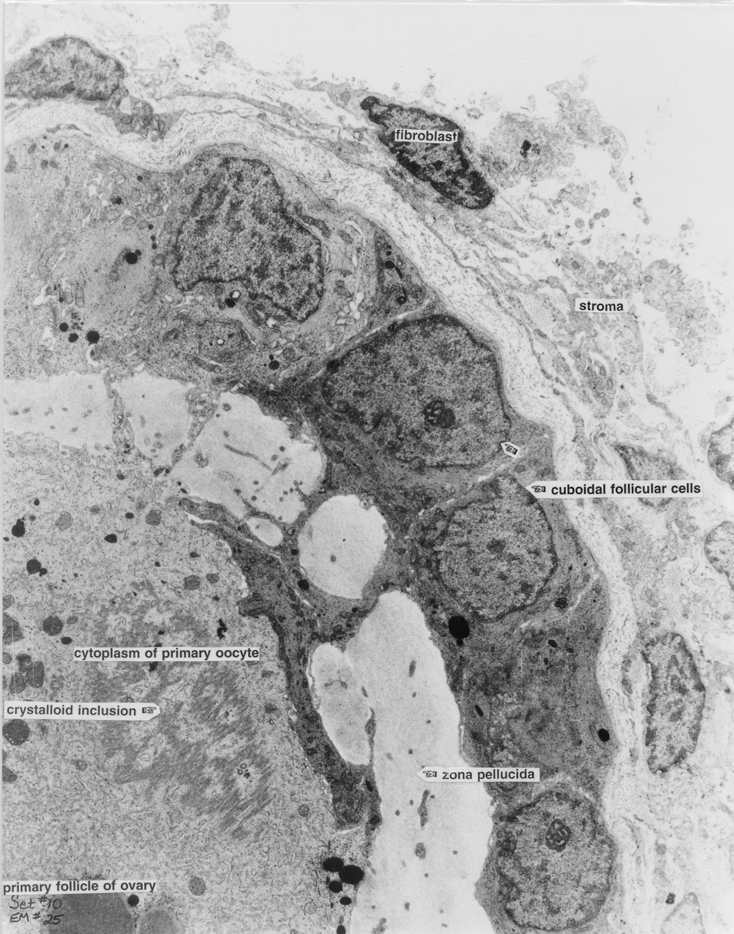
Ovary 25 |
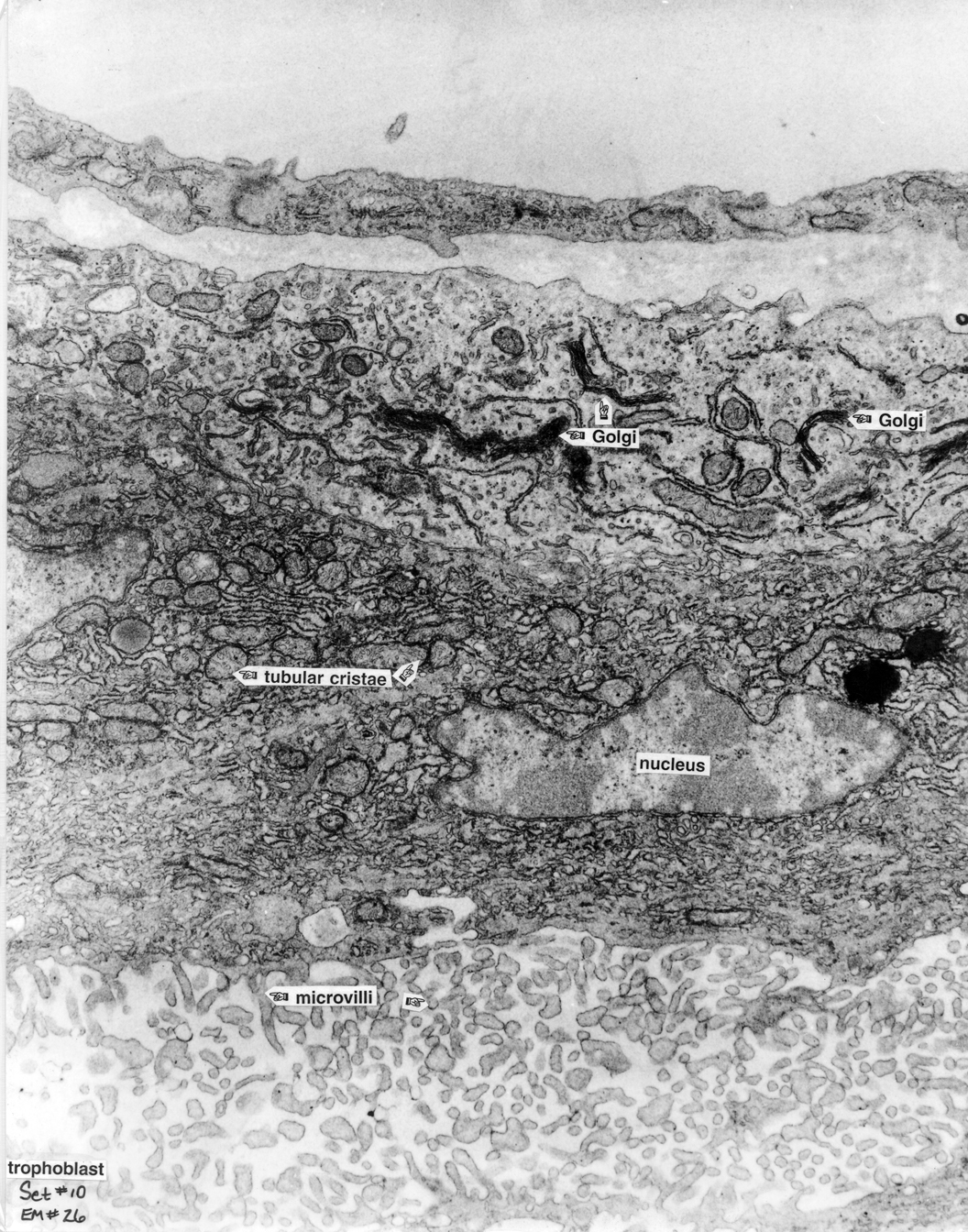
Fetal Tissue 26 |
||